

Common deactivation patterns during working memory and visual attention tasks: an intra-subject fMRI study at 4 Tesla
- PMID: 16404736
- PMCID: PMC2424317
- DOI: 10.1002/hbm.20211
Common deactivation patterns during working memory and visual attention tasks: an intra-subject fMRI study at 4 Tesla
Abstract
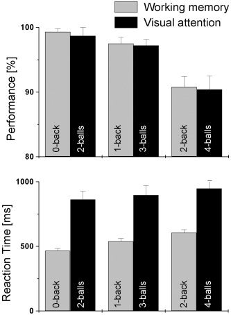
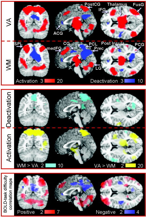
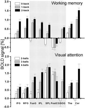
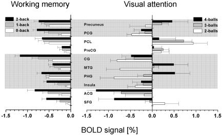
This parametric functional magnetic resonance imaging (fMRI) study investigates the balance of negative and positive fMRI signals in the brain. A set of visual attention (VA) and working memory (WM) tasks with graded levels of difficulty was used to deactivate separate but overlapping networks that include the frontal, temporal, occipital, and limbic lobes; regions commonly associated with auditory and emotional processing. Brain activation (% signal change and volume) was larger for VA tasks than for WM tasks, but deactivation was larger for WM tasks. Load-related increases of blood oxygenation level-dependent (BOLD) responses for different levels of task difficulty cross-correlated strongly in the deactivated network during VA but less so during WM. The variability of the deactivated network across different cognitive tasks supports the hypothesis that global cerebral blood flow vary across different tasks, but not between different levels of task difficulty of the same task. The task-dependent balance of activation and deactivation might allow maximization of resources for the activated network.
Figures
Similar articles
-
Specialization in the default mode: Task-induced brain deactivations dissociate between visual working memory and attention.Hum Brain Mapp. 2010 Jan;31(1):126-39. doi: 10.1002/hbm.20850. Hum Brain Mapp. 2010. PMID: 19639552 Free PMC article.
-
Different activation patterns for working memory load and visual attention load.Brain Res. 2007 Feb 9;1132(1):158-65. doi: 10.1016/j.brainres.2006.11.030. Epub 2006 Dec 12. Brain Res. 2007. PMID: 17169343 Free PMC article.
-
Crossmodal influences in somatosensory cortex: Interaction of vision and touch.Hum Brain Mapp. 2010 Jan;31(1):14-25. doi: 10.1002/hbm.20841. Hum Brain Mapp. 2010. PMID: 19572308 Free PMC article.
-
Discrete capacity limits in visual working memory.Curr Opin Neurobiol. 2010 Apr;20(2):177-82. doi: 10.1016/j.conb.2010.03.005. Epub 2010 Mar 31. Curr Opin Neurobiol. 2010. PMID: 20362427 Free PMC article. Review.
-
Representations of spectral coding in the human brain.Int Rev Neurobiol. 2005;70:331-69. doi: 10.1016/S0074-7742(05)70010-6. Int Rev Neurobiol. 2005. PMID: 16472639 Review. No abstract available.
Cited by
-
Altered Value Coding in the Ventromedial Prefrontal Cortex in Healthy Older Adults.Front Aging Neurosci. 2016 Aug 31;8:210. doi: 10.3389/fnagi.2016.00210. eCollection 2016. Front Aging Neurosci. 2016. PMID: 27630561 Free PMC article.
-
Disentangling linear and nonlinear brain responses to evoked deep tissue pain.Pain. 2012 Oct;153(10):2140-2151. doi: 10.1016/j.pain.2012.07.014. Epub 2012 Aug 9. Pain. 2012. PMID: 22883925 Free PMC article.
-
Disrupted functional connectivity with dopaminergic midbrain in cocaine abusers.PLoS One. 2010 May 25;5(5):e10815. doi: 10.1371/journal.pone.0010815. PLoS One. 2010. PMID: 20520835 Free PMC article.
-
How processing emotion affects language control in bilinguals.Brain Struct Funct. 2023 Mar;228(2):635-649. doi: 10.1007/s00429-022-02608-5. Epub 2022 Dec 31. Brain Struct Funct. 2023. PMID: 36585969
-
Brain structures and activity during a working memory task associated with internet addiction tendency in young adults: A large sample study.PLoS One. 2021 Nov 15;16(11):e0259259. doi: 10.1371/journal.pone.0259259. eCollection 2021. PLoS One. 2021. PMID: 34780490 Free PMC article.
References
-
- Aguirre GK, Zarahn E, D'Esposito M (1998): The inferential impact of global signal covariates in functional neuroimaging analyses. Neuroimage 8: 302–306. - PubMed
-
- Ashburner J, Neelin P, Collins DL, Evans AC, Friston KJ (1997): Incorporating prior knowledge into image registration. Neuroimage 6: 344–352. - PubMed
-
- Born AP, Law I, Lund TE, Rostrup E, Hanson LG, Wildschiodtz G, Lou HC, Paulson OB (2002): Cortical deactivation induced by visual stimulation in human slow‐wave sleep. Neuroimage 17: 1325–1335. - PubMed
-
- Brown S, Martinez M, Parsons L (2004): Passive music listening spontaneously engages limbic and paralimbic systems. Neuroreport 15: 2033–2037. - PubMed
-
- Buxton RB. 2002. Introduction to functional magnetic resonance imaging: principles and techniques. Cambridge: Cambridge University Press.
Publication types
MeSH terms
Grants and funding
LinkOut - more resources
Full Text Sources
Other Literature Sources
