. 2006 Jan;175(1):113-20.
doi: 10.1016/S0022-5347(05)00065-0.

Transrectal prostate biopsy and fiducial marker placement in a standard 1.5T magnetic resonance imaging scanner

Affiliations

Transrectal prostate biopsy and fiducial marker placement in a standard 1.5T magnetic resonance imaging scanner

Robert C Susil et al. J Urol. 2006 Jan.

Abstract

Purpose: We investigated the accuracy and feasibility of a system that provides transrectal needle access to the prostate concurrent with 1.5 Tesla MRI which previously has not been possible.

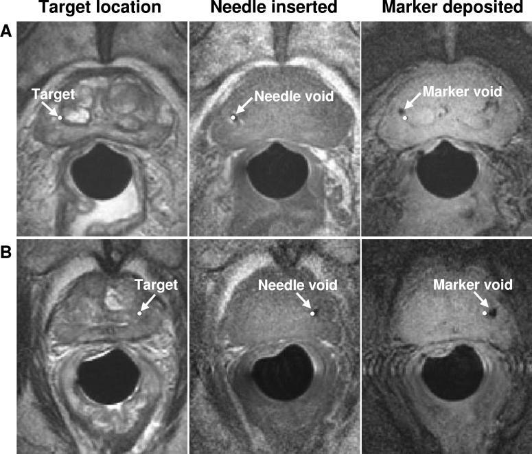
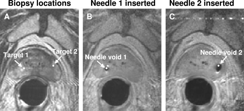
Materials and methods: In 5 patients with previously diagnosed prostate cancer, MRI guided intraprostatic placement of gold fiducial markers (4 procedures) and/or prostate biopsy (3 procedures) was performed using local anesthesia.

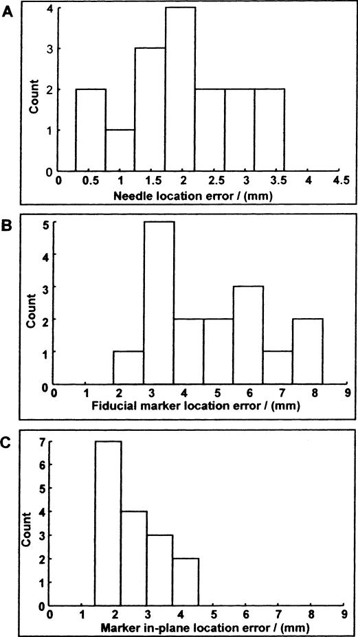
Results: Mean procedure duration was 76 minutes and all patients tolerated the intervention well. Procedure related adverse events included self-limited hematuria and hematochezia following 3 of 8 procedures (all resolved in less than 1 week). Mean needle placement accuracy was 1.9 mm for the fiducial marker placement studies and 1.8 mm for the biopsy procedures. Mean fiducial marker placement accuracy was 4.8 mm and the mean fiducial marker placement accuracy transverse to the needle direction was 2.6 mm. All patients who underwent the procedure were able to complete their course of radiotherapy without delay or complication.

Conclusions: While studies of clinical usefulness are warranted, transrectal 1.5 T MRI guided prostate biopsy and fiducial marker placement is feasible using this system, providing new opportunities for image guided diagnostic and therapeutic prostate interventions.


Figures

Fig. 1
Fig. 1
Interventional system. A, stationary endorectal sheath with integrated 20 mm diameter single turn imaging coil minimizes tissue deformation during interventional procedure. B, cylindrical needle guide contains MR tracking microcoils and curved needle channel. C, positioning stage houses mechanism that converts rotation of 2 control rods into rotation and translation of needle guide, which fits inside endorectal sheath. D, positioning arm with 2 lockable aluminum ball joints and linear rail allows for easy device positioning and, subsequently, rigid immobilization.
Fig. 2
Fig. 2
Prostate biopsy needle guide and sheath. A, to accommodate 14 gauge spring-loaded biopsy guns, modified needle guide with 2, straight needle channels (30 degrees and 20 degrees) and modified endorectal sheath were used. B, sagittal T1-weighted fast spin-echo image was acquired with biopsy needle in place. Using 1 of 2 needle channels, all sites within prostate could be accessed.
Fig. 3
Fig. 3
MR scanner room setup. A, flexible control rods (2) extend to edge of scanner bore, allowing for rotation and translation of needle guide while prostate is located at scanner isocenter. B, image display and targeting application is projected onto screen in scanner room.
Fig. 4
Fig. 4
Image display and targeting application. Target (white circle) is graphically selected within diffuse, low intensity region (corresponding to region cancerous tissue) in left posterior lobe of prostate. Intersection of image plane and projected needle path (white cross) as well as axis of needle guide (white square) are also shown. As device is moved, targeting commands and projected needle path are updated in real time.
Fig. 5
Fig. 5
Targeting, needle and fiducial-marker visualization images. Images from 2 patients (A and B, respectively), show images acquired during fiducial-marker placement procedure. Column 1, targets are selected on axial, T2-weighted FSE images. Column 2, needle tip void is visualized in axial, T1-weighted FSE images. Column 3, marker void is visualized on axial, T2*-weighted gradient-echo images. Note that there is minimal tissue motion throughout each procedure.
Fig. 6
Fig. 6
Needle and fiducial-marker placement accuracy shown by error histograms for all 16 gold fiducial markers placed. A, needle tip location errors. B, fiducial marker location errors. C, fiducial marker in-plane location errors. Mean placement errors for each are 1.9, 4.8 and 2.6 mm, respectively. Because tissue core biopsies are typically 1.5 cm long, last measure, fiducial marker in-plane placement error, is best predictor of tissue biopsy acquisition accuracy.
Fig. 7
Fig. 7
Biopsy target and needle visualization images. A, biopsy locations were selected on axial, T2-weighted fast spin-echo images. B and C, 14 gauge biopsy needle void is visualized in axial, T1-weighted fast spin-echo images.
Fig. 8
Fig. 8
T1-weighted axial fast-spin-echo images of prostate. A, acquired 20 minutes after start of procedure. B, 40 minutes after start of procedure. C, 50 minutes after start of procedure. Throughout intervention, including 4 needle placements, prostate and surrounding tissues remain stable. Importantly stationary endorectal sheath prevents movement of needle guide from causing tissue deformation.

Comment in

Similar articles

Cited by

References

    1. Yu KK, Hricak H. Imaging prostate cancer. Radiol Clin North Am. 2000;38:59. - PubMed
    1. Wefer AE, Hricak H, Vigneron DB, Coakley FV, Lu Y, Wefer J, et al. Sextant localization of prostate cancer: comparison of sextant biopsy, magnetic resonance imaging and magnetic resonance spectroscopic imaging with step section histology. J Urol. 2000;164:400. - PubMed
    1. Cormack RA, Kooy H, Tempany CM, D'Amico AV. A clinical method for real-time dosimetric guidance of transperineal 125I prostate implants using interventional magnetic resonance imaging. Int J Radiat Oncol Biol Phys. 2000;46:207. - PubMed
    1. D'Amico AV, Tempany CM, Cormack R, Hata N, Jinzaki M, Tuncali K, et al. Transperineal magnetic resonance image guided prostate biopsy. J Urol. 2000;164:385. - PubMed
    1. Cormack RA, D'Amico AV, Hata N, Silverman S, Weinstein M, Tempany CM. Feasibility of transperineal prostate biopsy under interventional magnetic resonance guidance. Urology. 2000;56:663. - PubMed

Publication types