. 2006 Jan 17;103(3):643-8.
doi: 10.1073/pnas.0507661103. Epub 2006 Jan 9.

Mammalian alpha I-spectrin is a neofunctionalized polypeptide adapted to small highly deformable erythrocytes

Affiliations

Mammalian alpha I-spectrin is a neofunctionalized polypeptide adapted to small highly deformable erythrocytes

Marcela Salomao et al. Proc Natl Acad Sci U S A. .

Abstract

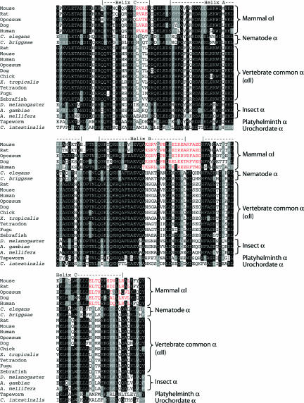
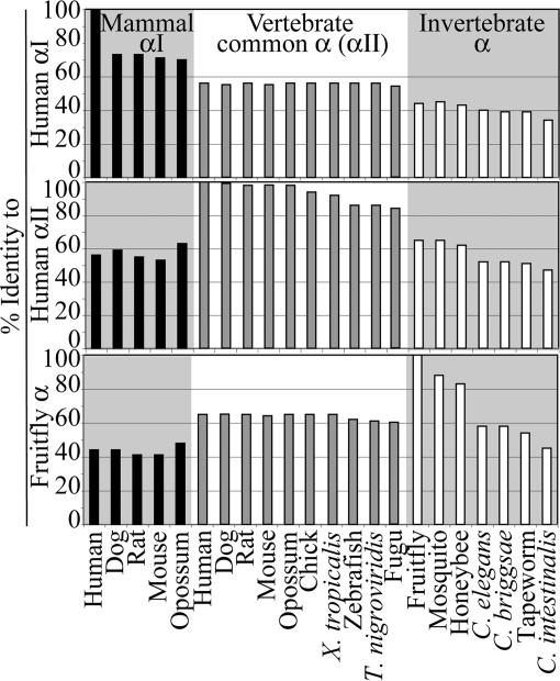
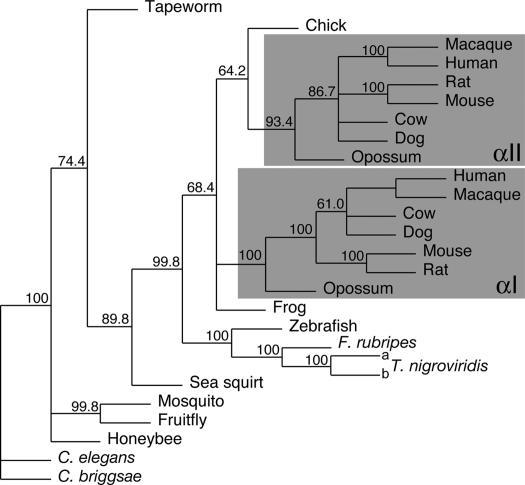
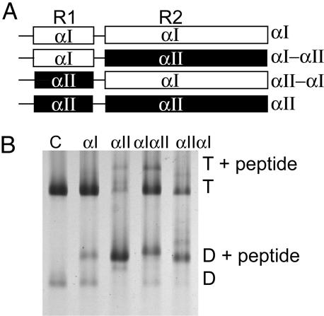
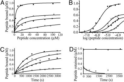
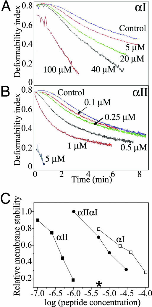
Mammalian red blood cells, unlike those of other vertebrates, must withstand the rigors of circulation in the absence of new protein synthesis. Key to this is plasma membrane elasticity deriving from the protein spectrin, which forms a network on the cytoplasmic face. Spectrin is a tetramer (alphabeta)(2), made up of alphabeta dimers linked head to head. We show here that one component of erythrocyte spectrin, alphaI, is encoded by a gene unique to mammals. Phylogenetic analysis suggests that the other alpha-spectrin gene (alphaII) common to all vertebrates was duplicated after the emergence of amphibia, and that the resulting alphaI gene was preserved only in mammals. The activities of alphaI and alphaII spectrins differ in the context of the human red cell membrane. An alphaI-spectrin fragment containing the site of head-to-head interaction with the beta-chain binds more weakly than the corresponding alphaII fragment to this site. The latter competes so strongly with endogenous alphaI as to cause destabilization of membranes at 100-fold lower concentration than the alphaI fragment. The efficacies of alphaI/alphaII chimeras indicate that the partial structural repeat, which binds to the complementary beta-spectrin element, and the adjacent complete repeat together determine the strength of the dimer-dimer interaction on the membrane. Alignment of all available alpha-spectrin N-terminal sequences reveals three blocks of sequence unique to alphaI. Furthermore, human alphaII-spectrin is closer to fruitfly alpha-spectrin than to human alphaI-spectrin, consistent with adaptation of alphaI to new functions. We conclude that alphaI-spectrin represents a neofunctionalized spectrin adapted to the rapid make and break of tetramers.


Figures

Fig. 1.
Fig. 1.
Alignment of the amino acid sequences of the N-terminal regions of α-spectrins. Shown are the alignment of a structurally well defined and functionally essential region of α-spectrin, namely the N-terminal helix that binds β-spectrin and the first full triple-helical repeat. The secondary structure elements are assigned by alignment with the NMR structure of human αI-spectrin (Protein Data Bank ID code 1OWA).
Fig. 2.
Fig. 2.
Sequence identities between αI-, αII-, and invertebrate α-spectrins. The sequences of the N-terminal regions of human αI-, αII-, or fruitfly α-spectrin were compared with other sequences, as indicated.
Fig. 3.
Fig. 3.
Maximum likelihood phylogenetic tree of α-spectrins. cDNA sequences encoding the region from the N-terminal helix through the second full repeat were aligned and analyzed by maximum likelihood with the aid of phyml. Five hundred bootstrapped replicates were examined, and a consensus tree was built from these by using phylip consense. Numbers shown at forks indicate the percentage occurrence among the trees of the group consisting of the species to the right of that fork. The groups of organisms containing both αI- and αII-spectrin are shaded. The organisms and cDNA sequences used are as listed in Table 1.
Fig. 4.
Fig. 4.
Binding of αI- and αII-spectrin fragments to spectrin dimer self-association sites in ghost membranes at 37°C. (A) Schematic diagram of α-spectrin fragments. R1 is the partial triple helical repeat that binds to β.R2 is the first full triple helix. αIαII and αIIαI are hybrids in which R1 and R2 are swapped between the proteins. (B) Spectrin extracted from the resealed ghosts was analyzed by electrophoresis in 5% nondenaturing gels. Incorporation at 100 μM total concentration of each fragment was demonstrated by the presence of a new band, migrating above the spectrin dimer (D) and tetramer (T). Lane 1, control spectrin with no peptide introduced into the ghosts; lane 2, αI fragment incorporated; lane 3, αII fragment incorporation; lane 4, αIαII; lane 5 αIIαI.
Fig. 5.
Fig. 5.
Binding isotherms and kinetics of interaction for α-spectrin fragments to spectrin in intact membranes. (A and B) Binding of α-spectrin fragments to membranes. Open squares, αI; solid squares, αII; open circles, αIIαI; solid circles, αIαII. The curves are calculated, assuming a set of independent identical sites in each case. B shows the data plotted on a log scale. (C) Association rates for αI (open squares), αII (filled circles), αIαII (filled squares), and αIIαI (open circles) peptides; (D) dissociation rates of αI (open circles) and αII (closed circles) peptides from membranes. The curve is calculated from the initial rate (see text).
Fig. 6.
Fig. 6.
Effect of peptide incorporation on membrane stability. Membrane mechanical stability of the resealed ghosts was measured by ektacytometry. Membrane stability is expressed in terms of the rate of decline in deformability index (DI). Faster decay (DI curve shifted to the left) reflects destabilization of the membrane. A and B show the effects of incorporation of the αI and αII peptides, respectively; (C) correlation between relative membrane stability (the time taken to reach 50% loss of deformability) and peptide concentrations; open squares, αI; filled squares, αII; filled circles, αIIαI. *, A concentration of αII peptide that caused essentially instantaneous fragmentation.

References

    1. Gulliver, G. (1875) Proc. Zool. Soc. London 1875, 474–495.
    1. Hawkey, C. M., Bennett, P. M., Gascoyne, S. C., Hart, M. G. & Kirkwood, J. K. (1991) Br. J. Haematol. 77, 392–397. - PubMed
    1. Gregory, T. R. (2001) Blood Cells Mol. Dis. 27, 830–843. - PubMed
    1. Mohandas, N. & Evans, E. (1994) Annu. Rev. Biophys. Biomol. Struct. 23, 787–818. - PubMed
    1. Discher, D. E. & Carl, P. (2001) Cell Mol. Biol. Lett. 6, 593–606. - PubMed

Publication types

LinkOut - more resources