. 2006 Jan 10;103(2):341-6.
doi: 10.1073/pnas.0506618103. Epub 2006 Jan 3.

The activity of a human endoplasmic reticulum-associated degradation E3, gp78, requires its Cue domain, RING finger, and an E2-binding site

Affiliations

The activity of a human endoplasmic reticulum-associated degradation E3, gp78, requires its Cue domain, RING finger, and an E2-binding site

Bo Chen et al. Proc Natl Acad Sci U S A. .

Abstract

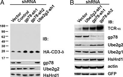
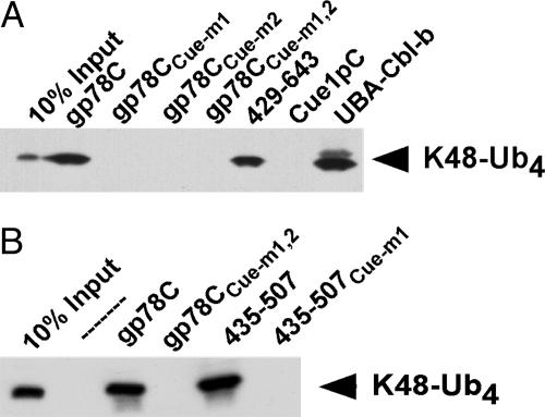
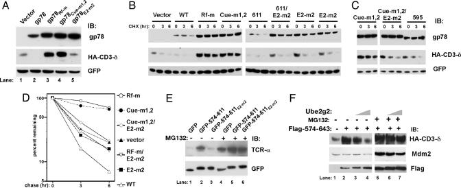
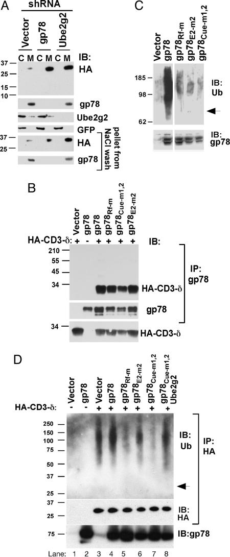
Efficient targeting of proteins for degradation from the secretory pathway is essential to homeostasis. This occurs through endoplasmic reticulum (ER)-associated degradation (ERAD). In this study, we establish that a human ubiquitin ligase (E3), gp78, and a specific E2, Ube2g2, are both critically important for ERAD of multiple substrates. gp78 exhibits a complex domain structure that, in addition to the RING finger, includes a ubiquitin-binding Cue domain and a specific binding site for Ube2g2. Disruption of either of these domains abolishes gp78-mediated ubiquitylation and protein degradation, resulting in accumulation of substrates in their fully glycosylated forms in the ER. This suggests that gp78-mediated ubiquitylation is an early step in ERAD that precedes dislocation of substrates from the ER. The in vivo requirement for both an E2-binding site distinct from the RING finger and a ubiquitin-binding domain intrinsic to an E3 suggests a previously unappreciated level of complexity in ubiquitin ligase function. These results also provide proof of principle that interrupting a specific E2-E3 interaction can selectively inhibit ERAD.


Figures

Fig. 1.
Fig. 1.
Essential roles for gp78/autocrine motility factor receptor (AMFR) and Ube2g2 in ERAD. (A) HT1080 cells were cotransfected with the indicated shRNA plasmids and HA-tagged CD3-δ. After 48 h, cell lysates were analyzed by immunoblotting (IB) after resolution of equal amounts of lysates on duplicate gels. (B) Human embryonic kidney 293T cells were cotransfected with shRNA plasmids and TCR-α. gp78 and TCR-α were first immunoprecipitated before SDS/PAGE. Transfection efficiency was monitored by GFP.
Fig. 2.
Fig. 2.
Localization and specificity of Ube2g2 binding to gp78. (A) Schematic representation of full-length gp78 showing RING finger (Rf) and Cue domain (Cue). (B) (Upper) Schematic of gp78 cytoplasmic domain with RING finger, Cue domain and minimal G2BR indicated. GST fusions tested for Ube2g2 binding are shown below with summary of results at right. (Lower) Alignment of gp78 G2BR with the analogous region of Cue1p. Mutations and their effects on E2 binding are shown below the gp78 sequence. (C) Ube2g2 was translated in reticulocyte lysate with 35S-Met and incubated with the indicated GST fusions bound to beads and assessed for binding. (D) Bacterially expressed Ube2g2 was reacted with E1 with or without ubiquitin and bound to the indicated GST fusions. Washed samples were eluted from beads in the absence (NR) or presence (R) of reducing agent (2-mercaptoethanol) before SDS/PAGE and IB with anti-Ube2g2. (E) Human embryonic kidney (HEK) 293T cells were cotransfected with Myc-Ube2g2 and gp78 or the indicated mutants. Rf-m is an inactivating RING finger mutation. Subscripts 595 and 611 are truncations after the indicated amino acids. E2-m2 corresponds to the mutation in the G2BR described in B. Samples were treated with MG132 for 8 h before lysis. Immunoprecipitation (IP) (Upper) with anti-Myc was followed by IB, as indicated. Lysate corresponding to 10% of the material used for IP was directly immunoblotted for gp78 (Lower). (F) HEK 293T were transfected with plasmid encoding the indicated amino acids of gp78 fused to the C terminus of GFP. Anti-GFP IPs (Left) or whole-cell lysates corresponding to ≈10% of the amount used for IP (Right) were immunoblotted with antibodies raised against the indicated E2s.
Fig. 3.
Fig. 3.
gp78 binds ubiquitin through its Cue domain. (A and B) Equal amounts of GST fusions were incubated with K48-Ub4 and binding determined by IB with antiubiquitin. gp78CCue-m1 includes the complete C-terminal region of gp78 with an MFP to GGR mutation of 467 to 469. gp78Cue-m2 contains a VLQDL to RLQVD mutation of 476 to 480. gp78CCue-m1,2 contains both sets of mutations. Cue1pC and UBA-Cbl-b correspond to GST fusions of the cytoplasmic domain of Cue1p and the UBA of Cbl-b, respectively. 435-507 and 435-507Cue-m1 are GST fusions of amino acids 435-507 of gp78 either without or with the Cue-m1 mutation.
Fig. 4.
Fig. 4.
Multiple domains of gp78 are required to target itself and a heterologous ERAD substrate for degradation. (A-C) Cells were cotransfected with the indicated forms of gp78 together with CD3-δ and lysates analyzed by IB. (B and C) Cells were treated with cycloheximide (CHX), as indicated. (D) Graphic representation of CD3-δ levels from B and C (full length gp78 transfections only). Cue-m1,2 is the average of data shown in B and C. (E) Cells were cotransfected with TCR-α and GFP fusions of gp78 and treated with MG132 where indicated. (F) Cells transfected with CD3-δ and Mdm2 were cotransfected with Flag-tagged amino acids 574-643 of gp78 where indicated. In lanes 3 and 6, 2 μg of Ube2g2 plasmid was cotransfected; in lanes 4 and 7, 4 μg was used. MG132 treatment was for 8 h.
Fig. 5.
Fig. 5.
The G2BR and Cue domain are required for in vivo E3 activity. (A) Membrane (M) and cytoplasmic (C) fractions from HT1080 cells transfected with HA-CD3-δ and the indicated shRNAs were prepared by hypotonic lysis and equal cell equivalents of each resolved by SDS/PAGE. Membrane fractions were subject to a high salt wash to further remove nonintegral membrane proteins. (B) Cells transfected as indicated were treated with MG132 for 5 h before IP with anti-gp78 followed by IB as indicated for gp78 or HA-CD3-δ. (Lower) IB of whole cell lysate representing 10% of the material used for IP. Similar co-IP was obtained when IPs were washed in either Triton X-100 buffer (shown) or RIPA buffer. (C) Cells were transfected with WT or mutant gp78 plasmids as indicated. Samples were treated with MG132 for 8 h and lysates immunoprecipitated with anti-gp78 followed by IB with either antiubiquitin (Upper) or anti-gp78 (Lower). Arrow indicates migration of unmodified gp78. (D) Cells were transfected with CD3-δ and gp78 as indicated. In lane 8, plasmid encoding Ube2g2 was coexpressed. Cells were treated with MG132 as in C before lysis and IP with anti-HA. Arrow indicates migration of CD3-δ.

Similar articles

Cited by

References

    1. Hampton, R. Y. (2002) Curr. Opin. Cell Biol. 14, 476-482. - PubMed
    1. Kostova, Z. & Wolf, D. H. (2003) EMBO J. 22, 2309-2317. - PMC - PubMed
    1. Jarosch, E., Lenk, U. & Sommer, T. (2003) Int. Rev. Cytol. 223, 39-81. - PubMed
    1. Bays, N. W., Gardner, R. G., Seelig, L. P., Joazeiro, C. A. & Hampton, R. Y. (2001) Nat. Cell Biol. 3, 24-29. - PubMed
    1. Deak, P. M. & Wolf, D. H. (2001) J. Biol. Chem. 276, 10663-11069. - PubMed

Publication types