. 2006 Jan 10;103(2):443-8.
doi: 10.1073/pnas.0503839103. Epub 2006 Jan 3.

Nonsteroidal anti-inflammatory drugs repress beta-secretase gene promoter activity by the activation of PPARgamma

Affiliations

Nonsteroidal anti-inflammatory drugs repress beta-secretase gene promoter activity by the activation of PPARgamma

Magdalena Sastre et al. Proc Natl Acad Sci U S A. .

Abstract

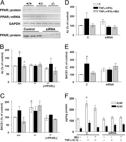
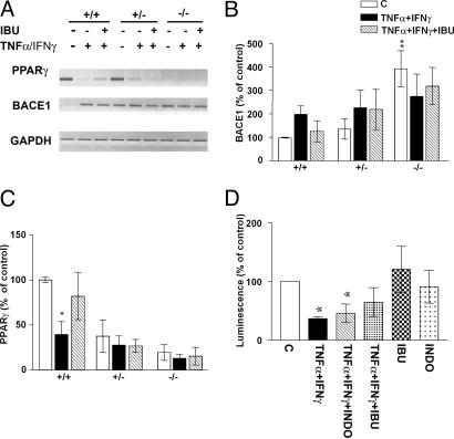
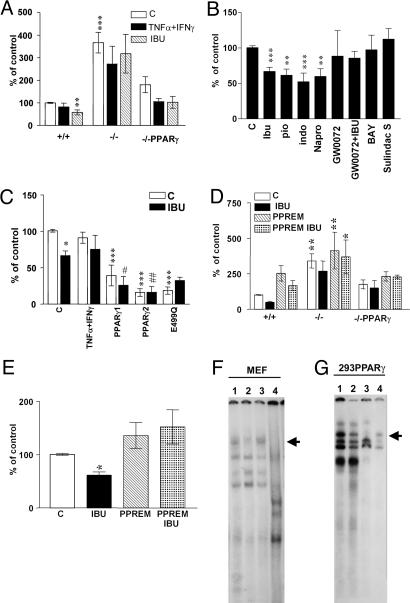
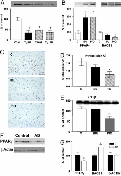
Epidemiological evidence suggests that nonsteroidal anti-inflammatory drugs (NSAIDs) decrease the risk for Alzheimer's disease (AD). Certain NSAIDs can activate the peroxisome proliferator-activated receptor-gamma (PPARgamma), which is a nuclear transcriptional regulator. Here we show that PPARgamma depletion potentiates beta-secretase [beta-site amyloid precursor protein cleaving enzyme (BACE1)] mRNA levels by increasing BACE1 gene promoter activity. Conversely, overexpression of PPARgamma, as well as NSAIDs and PPARgamma activators, reduced BACE1 gene promoter activity. These results suggested that PPARgamma could be a repressor of BACE1. We then identified a PPARgamma responsive element (PPRE) in the BACE1 gene promoter. Mutagenesis of the PPRE abolished the binding of PPARgamma to the PPRE and increased BACE1 gene promoter activity. Furthermore, proinflammatory cytokines decreased PPARgamma gene transcription, and this effect was supressed by NSAIDs. We also demonstrate that in vivo treatment with PPARgamma agonists increased PPARgamma and reduced BACE1 mRNA and intracellular beta-amyloid levels. Interestingly, brain extracts from AD patients showed decreased PPARgamma expression and binding to PPRE in the BACE1 gene promoter. Our data strongly support a major role of PPARgamma in the modulation of amyloid-beta generation by inflammation and suggest that the protective mechanism of NSAIDs in AD involves activation of PPARgamma and decreased BACE1 gene transcription.


Figures

Fig. 1.
Fig. 1.
APP metabolism in cells that do not express PPARγ. (A) PPARγ expression and mRNA representation of MEF wild-type cells, heterozygous and knockout for PPARγ. PPARγ expression in N2a-sw cells transfected with siRNA for PPARγ.(B) Quantification of Aβ levels (n = 5) in MEF cells nonstimulated, stimulated with IFN-γ (1 ng/ml) + TNF-α (30 ng/ml) overnight, and then incubated with ibuprofen (IBU) (10 μM) for 4 h. (C) Quantification of BACE1 protein expression in MEF knockout for PPARγ with the same conditions as above. (D) Quantification of Aβ levels (n = 5) in N2a-sw cells transfected with siRNA stimulated as above. (E) Quantification of BACE1 expression in N2a-sw cells with the same conditions as above. Columns represent mean ± SEM. (F) Analysis of Aβ1-40 and Aβ1-42 secretion in HEK293 APPsw cells transfected with siRNA for PPARγ by ELISA (n = 3). Asterisks, significant differences between control and treatment. #, significant differences between treatment with cytokines alone and with NSAIDs. *, P ≤ 0.05; #, P ≤ 0.05, ANOVA followed by a Tukey post hoc test.
Fig. 2.
Fig. 2.
Transcriptional regulation of PPARγ and BACE1 by inflammatory cytokines and NSAIDs. (A) Modulation of BACE1 and PPARγ steady-state mRNA levels by IFN-γ (1 ng/ml) + TNF-α (30 ng/ml) is reversed with ibuprofen (IBU) (10 μM) in wild-type but not in knockout cells, shown by semiquantitative RT-PCR analysis of BACE mRNA. (B) Quantification of BACE1 steady-state mRNA levels in four experiments performed in PPARγ wild-type, heterozygous, and knockout cells. (C) Quantification of PPARγ steady-state mRNA levels in four experiments performed with the same samples. Columns represent mean ± SEM. (D) N2a-Sw cells transfected with PPARγ1 luciferase reporter construct were stimulated with IFN-γ (1 ng/ml) + TNF-α (30 ng/ml) with or without IBU (10 μM) or INDO(10 μM) (n = 3). *, P ≤ 0.05 ANOVA followed by a Tukey post hoc test.
Fig. 3.
Fig. 3.
PPARγ modulates BACE1 promoter activity. (A) Luciferase activities of a 1.5-kb BACE1 promoter/luciferase reporter construct transfected in PPARγ wild-type, knockout, and knockout transfected with PPARγ cDNA MEF. Cells were treated with by IFN-γ (1 ng/ml) + TNF-α (30 ng/ml) with or without ibuprofen (IBU) (10 μM), n = 5. (B) NSAIDs and PPARγ agonists inhibit transcription of BACE1 promoter. N2a-sw cells transfected with BACE1 promoter and incubated with IBU, Pio, Indo, Napro, GW0072X (1 μM), BAY11-7082, and sulindac sulfide n = 4, all at 10 μM concentration. (C) PPARγ transfection inhibits BACE1 promoter activity. N2a-sw cells were transfected with BACE1 promoter construct and PPARγ1, PPARγ2, and PPARγ2 E499Q cDNA and incubated with or without IBU (10 μM); n = 4. (D) MEF transfected with BACE1 promoter control and mutated at the PPRE site were incubated with IBU (10 μM). (E) N2a cells transfected with BACE1 promoter control and mutated at the PPRE site were incubated with IBU (10 μM). Columns represent mean ± SEM, n = 4. Asterisks, significant differences between wild-type cells and treated cells. #, differences between transfected cells treated or untreated. *, P ≤ 0.05; **, P ≤ 0.01; ***, P ≤ 0.001; #, P ≤ 0.05; ##, P ≤ 0.01, ANOVA followed by a Tukey post hoc test. (F) Gel-shift analysis with the BACE1-PPRE probe using nuclear extract from MEF cells. The major PPARγ-containing complex is indicated by the arrow. Lane 1, MEF wild-type cells; lane 2, MEF PPARγ knockout cells; lane 3, MEF PPARγ knockout cells transfected with PPARγ1 cDNA; lane 4, MEF wild-type cells incubated with a excess of unlabeled BACE1-PPRE probe. (G) Gel shift using HEK293 cells transfected with human PPARγ2 cDNA. Lane 1, control; lane 2, supershift analysis; lane 3, molar excess of unlabeled BACE1-PPRE probe; lane 4, labeled mutant BACE1-PPRE probe (BACE1-PPREM).
Fig. 4.
Fig. 4.
PPARγ in the brain of APPV717I transgenic mice and in AD patients. (A) Quantification of PPARγ expression in cortex from mouse brain control and APPV717I of 3 and 16 months of age. (B) Quantification of the mRNA levels of PPARγ and BACE1 APPV717I mice treated with ibuprofen (IBU) and pioglitazone (Pio). (C) Immunostaining of intracellular Aβ in subiculum from APPV717I mice treated with IBU and Pio. (D) Quantification of percentage of intracellular Aβ in subiculum from APPV717I mice treated with IBU and Pio. Bar, 100 μM. Columns represent mean ± SEM, n = 4. (E) Representative Western blot for CTF-β fragments in cortex from APPV717I mice treated with IBU and Pio. (F) Representative Western blot for PPARγ expression in brain lysates from two control and two AD patients. As a negative control, β-actin expression was analyzed. (G) Quantification of the levels of β-actin, PPARγ, and BACE1 in the frontal cortex of 10 controls and 10 AD patients. Asterisks, significant differences between control and AD. *, P ≤ 0.05; **, P ≤ 0.01. ANOVA followed by a Tukey post hoc test or Student's t test for human brains.

Similar articles

Cited by

References

    1. Vassar, R., Bennett, B. D., Babu-Khan, S., Kahn, S., Mendiaz, E. A., Denis, P., Teplow, D. B., Ross, S, Amarante, P., Loeloff, R., et al. (1999) Science 286, 735-741. - PubMed
    1. Luo, Y., Bolon, B., Damore, M. A., Fitzpatrick, D., Liu, H., Zhang, J., Yan, Q., Vassar, R. & Citron, M. (2003) Neurobiol. Dis. 14, 81-88. - PubMed
    1. Ohno, M., Sametsky, E. A., Younkin, L. H., Oakley, H., Younkin, S. G., Citron, M., Vassar, R. & Disterhoft, J. F. (2004) Neuron 41, 27-33. - PubMed
    1. Tamagno, E., Bardini, P., Obbili, A., Vitali, A., Borghi, R., Zaccheo, D., Pronzato, M. A., Danni, O., Smith, M. A., Perry, G. & Tabato, M. (2002) Neurobiol. Dis. 10, 279-288. - PubMed
    1. Hartlage-Rubsamen, M., Zeitschel, U., Apelt, J., Gartner, U., Franke, H., Stahl, T., Gunther, A., Schliebs, R., Penkowa, M., Bigl, V. & Rossner, S. (2003) Glia 4, 169-179. - PubMed

Publication types

MeSH terms