

PDE1B2 regulates cGMP and a subset of the phenotypic characteristics acquired upon macrophage differentiation from a monocyte
- PMID: 16407168
- PMCID: PMC1326187
- DOI: 10.1073/pnas.0509972102
PDE1B2 regulates cGMP and a subset of the phenotypic characteristics acquired upon macrophage differentiation from a monocyte
Abstract
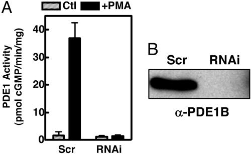
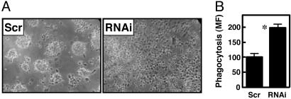
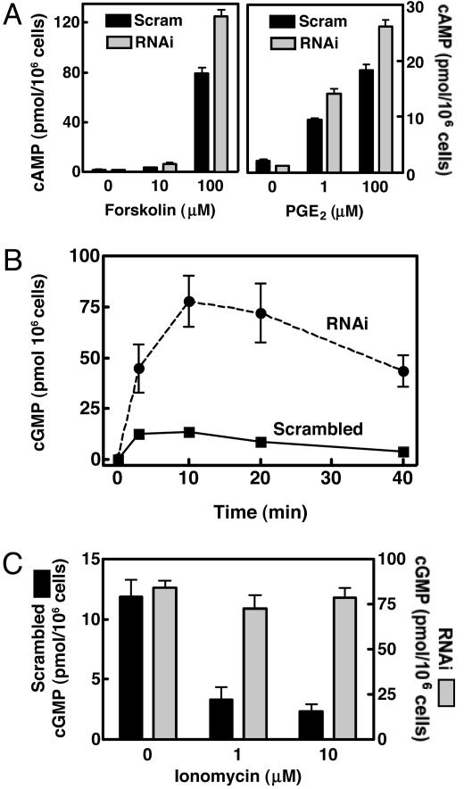
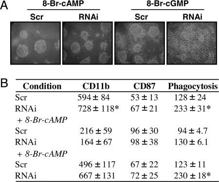
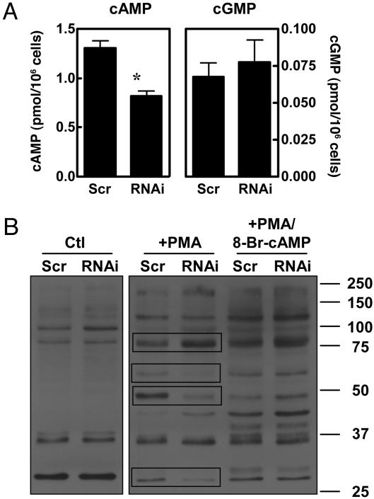
Monocyte-to-macrophage differentiation with the cytokine granulocyte-macrophage colony-stimulating factor induces expression of the cyclic nucleotide phosphodiesterase PDE1B2. However, what role PDE1B2 plays in macrophage biology has not been elucidated. We have addressed this question by inhibiting PDE1B2 induction by using RNA interference. Using a retrovirus-based system, we created HL-60 stable cell lines that express a short-hairpin RNA targeting PDE1B2. HL-60 cells treated with phorbol-12-myristate-13-acetate differentiate to a macrophage-like phenotype and up-regulate PDE1B2. However, expression of PDE1B2 short hairpin RNA effectively suppresses PDE1B2 mRNA, protein, and activity up-regulation. Using the HL-60 PDE1B2 knockdown cells and agonists for either adenylyl or guanylyl cyclase, it was found that PDE1B2 predominantly regulates cGMP and plays a lesser role in cAMP regulation in response to cyclase agonists. Furthermore, in intact HL-60 cells, PDE1B2 activity can be regulated by changes in Ca+2 levels. Inhibiting PDE1B2 up-regulation does not prevent HL-60 cell differentiation, because several markers of macrophage differentiation are unaffected. However, suppression of PDE1B2 expression alters some aspects of the macrophage-like phenotype, because cell spreading, phagocytic ability, and CD11b expression are augmented. The cAMP analog 8-Bromo-cAMP reverses the changes caused by PDE1B2 knockdown. Also, PDE1B2 knockdown cells have lower basal levels of cAMP and alterations in the phosphorylation state of several probable PKA substrate proteins. Thus, the effects of PDE1B2 on differentiation may ultimately be mediated through decreased cAMP. In conclusion, PDE1B2 regulates a subset of phenotypic changes that occur upon phorbol-12-myristate-13-acetate-induced differentiation and likely also plays a role in differentiated macrophages by regulating agonist-stimulated cGMP levels.
Figures
Similar articles
-
Cyclic nucleotides and phosphodiesterases in monocytic differentiation.Handb Exp Pharmacol. 2011;(204):365-90. doi: 10.1007/978-3-642-17969-3_16. Handb Exp Pharmacol. 2011. PMID: 21695649 Free PMC article. Review.
-
Selective up-regulation of PDE1B2 upon monocyte-to-macrophage differentiation.Proc Natl Acad Sci U S A. 2005 Jan 11;102(2):497-502. doi: 10.1073/pnas.0408535102. Epub 2004 Dec 29. Proc Natl Acad Sci U S A. 2005. PMID: 15625104 Free PMC article.
-
Differentiation of human monocytes in vitro with granulocyte-macrophage colony-stimulating factor and macrophage colony-stimulating factor produces distinct changes in cGMP phosphodiesterase expression.Cell Signal. 2004 Mar;16(3):365-74. doi: 10.1016/j.cellsig.2003.08.009. Cell Signal. 2004. PMID: 14687666
-
Characterization of cyclic nucleotide metabolism during human monocyte differentiation.J Leukoc Biol. 1984 Jun;35(6):617-30. doi: 10.1002/jlb.35.6.617. J Leukoc Biol. 1984. PMID: 6144716
-
Specific localized expression of cGMP PDEs in Purkinje neurons and macrophages.Neurochem Int. 2004 Nov;45(6):853-7. doi: 10.1016/j.neuint.2004.03.015. Neurochem Int. 2004. PMID: 15312979 Review.
Cited by
-
Ca2+/calmodulin-stimulated PDE1 regulates the beta-catenin/TCF signaling through PP2A B56 gamma subunit in proliferating vascular smooth muscle cells.FEBS J. 2010 Dec;277(24):5026-39. doi: 10.1111/j.1742-4658.2010.07908.x. Epub 2010 Nov 16. FEBS J. 2010. PMID: 21078118 Free PMC article.
-
Targeting cyclic nucleotide phosphodiesterase in the heart: therapeutic implications.J Cardiovasc Transl Res. 2010 Oct;3(5):507-15. doi: 10.1007/s12265-010-9203-9. Epub 2010 Jul 15. J Cardiovasc Transl Res. 2010. PMID: 20632220 Free PMC article. Review.
-
cGMP-dependent protein kinases and cGMP phosphodiesterases in nitric oxide and cGMP action.Pharmacol Rev. 2010 Sep;62(3):525-63. doi: 10.1124/pr.110.002907. Pharmacol Rev. 2010. PMID: 20716671 Free PMC article. Review.
-
Cyclic nucleotides and phosphodiesterases in monocytic differentiation.Handb Exp Pharmacol. 2011;(204):365-90. doi: 10.1007/978-3-642-17969-3_16. Handb Exp Pharmacol. 2011. PMID: 21695649 Free PMC article. Review.
-
Whole blood gene expression in adolescent chronic fatigue syndrome: an exploratory cross-sectional study suggesting altered B cell differentiation and survival.J Transl Med. 2017 May 11;15(1):102. doi: 10.1186/s12967-017-1201-0. J Transl Med. 2017. PMID: 28494812 Free PMC article.
References
Publication types
MeSH terms
Substances
Grants and funding
LinkOut - more resources
Full Text Sources
Research Materials
