. 2006 Mar 10;281(10):6691-8.
doi: 10.1074/jbc.M508314200. Epub 2006 Jan 1.

Group V secretory phospholipase A2 translocates to the phagosome after zymosan stimulation of mouse peritoneal macrophages and regulates phagocytosis

Affiliations

Group V secretory phospholipase A2 translocates to the phagosome after zymosan stimulation of mouse peritoneal macrophages and regulates phagocytosis

Barbara Balestrieri et al. J Biol Chem. .

Abstract

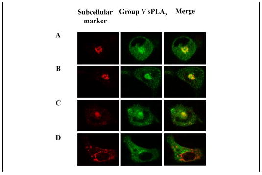
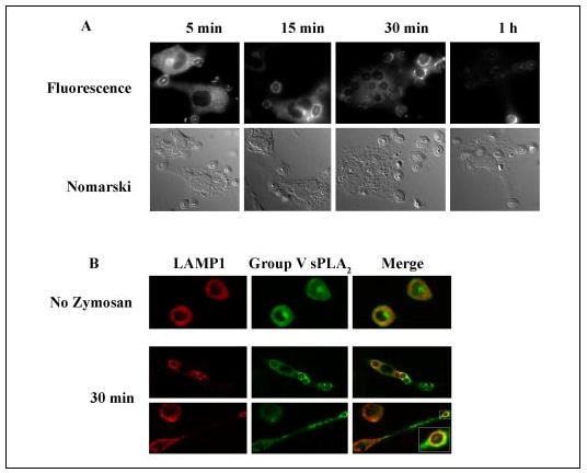
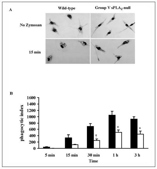
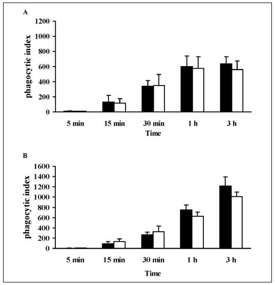
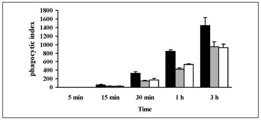
We have previously reported that group V secretory phospholipase A2 (sPLA2) amplifies the action of cytosolic phospholipase A2(cPLA2) alpha in regulating eicosanoid biosynthesis by mouse peritoneal macrophages stimulated with zymosan (Satake, Y., Diaz, B. L., Balestrieri, B., Lam, B. K., Kanaoka, Y., Grusby, M. J., and Arm, J. P. (2004) J. Biol. Chem. 279, 16488-16494). To further understand the role of group V sPLA2, we studied its localization in resting mouse peritoneal macrophages before and after stimulation with zymosan and the effect of deletion of the gene encoding group V sPLA2 on phagocytosis of zymosan. We report that group V sPLA2 is present in the Golgi apparatus and recycling endosome in the juxtanuclear region of resting peritoneal macrophages. Upon ingestion of zymosan by mouse peritoneal macrophages, group V sPLA2 is recruited to the phagosome. There it co-localizes with cPLA2alpha, 5-lipoxygenase, 5-lipoxygenase-activating protein, and leukotriene C4 synthase. Using immunostaining for the cysteinyl leukotrienes in carbodiimide-fixed cells, we show, for the first time, that the phagosome is a site of cysteinyl leukotriene formation. Furthermore, peritoneal macrophages from group V sPLA2-null mice demonstrated a >50% attenuation in phagocytosis of zymosan particles, which was restored by adenoviral expression of group V sPLA2 but IIA not group sPLA2. These data demonstrate that group V sPLA2 contributes to the innate immune response both through regulation of eicosanoid generation in response to a phagocytic stimulus and also as a component of the phagocytic machinery.


Figures

FIGURE 1
FIGURE 1. Subcellular localization of group V sPLA2
Peritoneal macrophages isolated from BALC/c mice were fixed with 2% paraformaldehyde, permeabilized with 0.025% saponin, and incubated with rabbit anti-group V sPLA2 and mAbs to GS28 (A), Syntaxin 6 (B), the transferrin receptor (C), or protein-disulfide isomerase (D). The cells were washed and incubated with FITC-conjugated donkey anti-rabbit IgG and Cy3-conjugated donkey anti-mouse IgG. Confocal images were obtained as described under “Experimental Procedures. ” Single color images are shown for the subcellular marker (Cy3, left panels) and group V sPLA2 (FITC, middle panels). The right panels are the merged images for both FITC and Cy3 staining.
FIGURE 2
FIGURE 2. Translocation of group V sPLA2 to the phagosome
A, peritoneal macrophages isolated from BALB/c mice were stimulated with unopsonized zymosan (10 particles/cell) for 5, 15, 30, or 60 min, fixed in 2% paraformaldehyde, permeabilized with 0.025% saponin, and incubated with rabbit anti-group V sPLA2 followed by FITC-conjugated donkey anti-rabbit IgG. Immunofluorescence images are paired with the corresponding Nomarski image. B, peritoneal macrophages isolated from BALB/c mice were left unstimulated or were stimulated with unopsonized zymosan (10 particles/cell) for 30 min, fixed in 2% paraformaldehyde, permeabilized with 0.025% saponin, and incubated with rabbit anti-group V sPLA2 and mAb to LAMP-1. The cells were washed and incubated with FITC-conjugated donkey anti-rabbit IgG and Cy3-conjugated donkey anti-rat IgG. Confocal images were obtained as described under “Experimental Procedures. ” Autofluorescence of zymosan particles is visible within each phagosome.
FIGURE 3
FIGURE 3. The phagosome is a site of LTC4 biosynthesis
A, kinetics of LTC4 generation by mouse peritoneal macrophages stimulated with zymosan (10 particles/cell). B, peritoneal macrophages isolated from BALB/c mice were stimulated with unopsonized zymosan (10 particles/cell) for 15 and 60 min, fixed in 2% paraformaldehyde, permeabilized with 0.025% saponin, and incubated with specific antibodies for each of the enzymes of LTC4 biosynthesis followed by FITC-conjugated secondary antibodies. C, to demonstrate LTC4 formation at the phagosome, the cells were fixed with EDAC and immunostained with rat mAb anti-Cys-LTs followed by Cy3-conjugated donkey anti-rat IgG. Immunofluorescence images are paired with the corresponding Nomarski or bright field image.
FIGURE 4
FIGURE 4. Phagocytosis of zymosan by group V sPLA2-null macrophages
A, light microscopy image of peritoneal macrophages derived from group V sPLA2-null mice (BALB/c N3) and wild-type littermates stained with Diff-Quick before and 15 min after the addition of zymosan (10 particles/cell). B, peritoneal macrophages from group V sPLA2- null mice (open bars) and from their wild-type littermate controls (filled bars) bred to a BALB/c background for three generations were incubated with 10 particles of zymosan/cell for 5 min to 3 h and stained with Diffquick. The phagocytic index was calculated as described under “Experimental Procedures. ” †, p < 0.02; *, p < 0.05 using Student’s paired t test.
FIGURE 5
FIGURE 5. Phagocytosis of zymosan by macrophages lacking cPLA2α (A) or LTC4 synthase (B)
Peritoneal macrophages from cPLA2α-null mice (A, open bars) and LTC4 synthase-null mice (B, open bars) and from their wild-type littermate controls (filled bars) were incubated with 10 particles of zymosan/cell for 5 min to 3 h and stained with Diffquick. The phagocytic index was calculated as described under “Experimental Procedures.”
FIGURE 6
FIGURE 6. Effects on phagocytosis of expressing group V sPLA2 and group IIA sPLA2 in group V sPLA2-null peritoneal macrophages
Peritoneal macrophages obtained from group V sPLA2-null mice bred to a BALB/c background (N10) were infected with adenovirus carrying group V sPLA2 (black bars), group IIA sPLA2 (gray bars), or LacZ (open bars) and GFP. After 48 h of infection cells were washed, stimulated with 10 particles/cell of zymosan for 5 min to 3 h and fixed with 2% paraformaldehyde. The cells were imaged as described under “Experimental Procedures, ” and the phagocytic index was calculated for GFP-positive cells

Similar articles

Cited by

References

    1. Soberman RJ, Christmas P. J Clin Investig. 2003;111:1107–1113. - PMC - PubMed
    1. Keller H, Dreyer C, Medin J, Mahfoudi A, Ozato K, Wahli W. Proc Natl Acad Sci U S A. 1993;90:2160–2164. - PMC - PubMed
    1. Zhu K, Baudhuin LM, Hong G, Williams FS, Cristina KL, Kabarowski JHS, Witte ON, Xu Y. J Biol Chem. 2001;276:41325–41335. - PubMed
    1. Clark JD, Lin LL, Kriz RW, Ramesha CS, Sultzman LA, Lin AY, Milona N, Knopf JL. Cell. 1991;65:1043–1051. - PubMed
    1. Pickard RT, Strifler BA, Kramer RM, Sharp JD. J Biol Chem. 1999;274:8823–8831. - PubMed

Publication types

MeSH terms