. 2006 Jan 10;34(1):89-95.
doi: 10.1093/nar/gkj416. Print 2006.

APOBEC3A and APOBEC3B are potent inhibitors of LTR-retrotransposon function in human cells

Affiliations

APOBEC3A and APOBEC3B are potent inhibitors of LTR-retrotransposon function in human cells

Hal P Bogerd et al. Nucleic Acids Res. .

Abstract

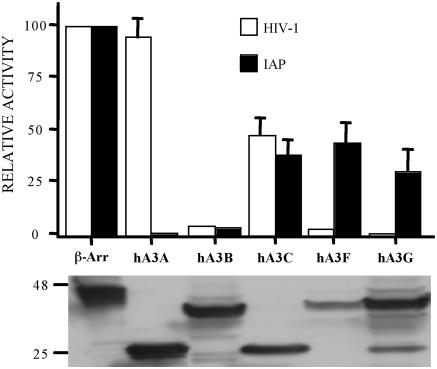
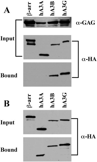
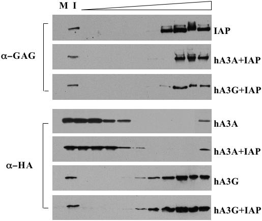
While the ability of APOBEC3G to reduce the replication of a range of exogenous retroviruses is now well established, recent evidence has suggested that APOBEC3G can also inhibit the replication of endogenous retrotransposons that bear long terminal repeats. Here, we extend this earlier work by showing that two other members of the human APOBEC3 protein family, APOBEC3B and APOBEC3A, can reduce retrotransposition by the intracisternal A-particle (IAP) retrotransposon in human cells by 20-fold to up to 100-fold, respectively. This compares to an approximately 4-fold inhibition in IAP retrotransposition induced by APOBEC3G. While both APOBEC3G and APOBEC3B specifically interact with the IAP Gag protein in co-expressing cells, and induce extensive editing of IAP reverse transcripts, APOBEC3A fails to package detectably into IAP virus-like particles and does not edit IAP reverse transcripts. These data, which identify human APOBEC3A as a highly potent inhibitor of LTR-retrotransposon function, are the first to ascribe a biological activity to APOBEC3A. Moreover, these results argue that APOBEC3A inhibits IAP retrotransposition via a novel mechanism that is distinct from, and in this case more effective than, the DNA editing mechanism characteristic of APOBEC3G and APOBEC3B.


Figures

Figure 1
Figure 1
Effect of human APOBEC3 proteins on IAP retrotransposition and HIV-1 infectivity. Upper panel: IAP retrotransposition was quantified by co-transfection of HeLa cells with the IAP retrotransposition indicator construct pDJ33/440N1neoTNF and the indicated APOBEC3 expression plasmids. After selection in G418, resistant colonies were counted. The effect of the same APOBEC3 proteins on HIV-1ΔVif infectivity was determined by co-transfection into 293T cells together with the pHIV-Luc-ΔVif indicator construct. Released HIV-1 virions were collected at 48 h and used to infect CD4+, CCR5+ cells. Induced, virus encoded luciferase activity was quantified 24 h later. In both cases, data are presented as a percentage of the activity seen in cultures co-transfected with a plasmid expressing the irrelevant β-arrestin gene. Average of three independent experiments with standard deviation indicated. Lower panel: western analysis of APOBEC3 protein expression in transfected 293T cells using an anti-HA tag specific mouse monoclonal. hA3A and hA3C are predicted to be ∼22 kDa in size while hA3B, hA3F and hA3G are ∼40 kDa.
Figure 2
Figure 2
Effect of APOBEC3 proteins on IAP retrotransposition and cell viability. (A) HeLa cells were transfected with the IAP retrotransposition indicator plasmid pDJ33/440N1neoTNF, together with an APOBEC3 expression plasmid or a control plasmid expressing β-arrestin. A derivative of pDJ33/440N1neoTNF lacking a functional pol gene served as the negative control. Cells were subjected to selection in G418 and resistant colonies counted 17 days after transfection. This experiment is representative of the data compiled in Figure 1. (B) Same as (A) except that HeLa cells were co-transfected with pcDNA3, which encodes a neo cDNA, instead of with pDJ33/440N1neoTNF. The negative in this case is a mock transfection.
Figure 3
Figure 3
An hA3A mutant bearing a defective cytidine deaminase active site remains able to inhibit IAP retrotransposition. These data were derived and are presented as described in Figure 1 and again used a primary antibody specific for the HA epitope tag, present on both the β-arrestin and hA3A proteins, in the lower panel. The hA3A (SPC-AAA) mutant bears alanine residues in place of the active site residues 99-SPC-101.
Figure 4
Figure 4
Binding of IAP Gag by APOBEC3 proteins in vivo and in vitro. (A) 293T cells were co-transfected with the IAP Gag expression plasmid pDJ33/440N1neoTNF and vectors expressing HA-tagged hA3A, hA3B, hA3G or β-arrestin, the latter as a negative control. After 48 h, the cells were lysed and an aliquot retained to analyze input protein levels. The remaining lysate was incubated with a rabbit anti-IAP Gag antiserum and protein A agarose. Bound, as well as input, proteins were separated by gel electrophoresis and visualized by western blot using a mouse monoclonal specific for the HA tag present on the APOBEC3 and β-arrestin proteins, or using the anti-IAP Gag antiserum. (B) 293T cells were transfected with plasmids expressing HA-tagged hA3A, hA3B, hA3G or β-arrestin and lysates prepared 2 days later. After incubation in the presence of purified full-length recombinant IAP Gag, Gag protein complexes were collected using a rabbit polyclonal anti-Gag antiserum and protein A agarose. Bound and input proteins were then visualized by western blot using an anti-HA monoclonal. For both panels, input lanes contain 2% of the starting material while bound lanes contain 25% of the immunoprecipitated protein fraction.
Figure 5
Figure 5
Sucrose gradient fractionation of IAP Gag and APOBEC3 proteins. 293T cells were transfected with pDJ33/440N1neoTNF, which expresses IAP Gag, with vectors expressing hA3G or hA3A, or with appropriate control plasmids. After 48 h, the cells were lysed and the lysates fractionated across 10–80% sucrose gradients. Fractions were then collected, with the top of the gradient at left, and subjected to western analysis using anti-IAP Gag and anti-HA tag antibodies, as indicated. M, mock transfected cell lysate; I, input cell lysate (0.5% of total).

Similar articles

Cited by

References

    1. Sheehy A.M., Gaddis N.C., Choi J.D., Malim M.H. Isolation of a human gene that inhibits HIV-1 infection and is suppressed by the viral Vif protein. Nature. 2002;418:646–650. - PubMed
    1. Wiegand H.L., Doehle B.P., Bogerd H.P., Cullen B.R. second human antiretroviral factor, APOBEC3F, is suppressed by the HIV-1 and HIV-2 Vif proteins. EMBO J. 2004;23:2451–2458. - PMC - PubMed
    1. Liddament M.T., Brown W.L., Schumacher A.J., Harris R.S. APOBEC3F properties and hypermutation preferences indicate activity against HIV-1 in vivo. Curr. Biol. 2004;14:1385–1391. - PubMed
    1. Zheng Y.-H., Irwin D., Kurosu T., Tokunaga K., Sata T., Peterlin B.M. Human APOBEC3F is another host factor that blocks human immunodeficiency virus type 1 replication. J. Virol. 2004;78:6073–6076. - PMC - PubMed
    1. Bishop K.N., Holmes R.K., Sheehy A.M., Davidson N.O., Cho S.-J., Malim M.H. Cytidine deamination of retroviral DNA by diverse APOBEC proteins. Curr. Biol. 2004;14:1392–1396. - PubMed

Publication types