. 2006 Mar;17(3):1410-20.
doi: 10.1091/mbc.e05-11-1005. Epub 2006 Jan 11.

PGAP2 is essential for correct processing and stable expression of GPI-anchored proteins

Affiliations

PGAP2 is essential for correct processing and stable expression of GPI-anchored proteins

Yuko Tashima et al. Mol Biol Cell. 2006 Mar.

Abstract

Biosynthesis of glycosylphosphatidylinositol-anchored proteins (GPI-APs) in the ER has been extensively studied, whereas the molecular events during the transport of GPI-APs from the ER to the cell surface are poorly understood. Here, we established new mutant cell lines whose surface expressions of GPI-APs were greatly decreased despite normal biosynthesis of GPI-APs in the ER. We identified a gene responsible for this defect, designated PGAP2 (for Post-GPI-Attachment to Proteins 2), which encoded a Golgi/ER-resident membrane protein. The low surface expression of GPI-APs was due to their secretion into the culture medium. GPI-APs were modified/cleaved by two reaction steps in the mutant cells. First, the GPI anchor was converted to lyso-GPI before exiting the trans-Golgi network. Second, lyso-GPI-APs were cleaved by a phospholipase D after transport to the plasma membrane. Therefore, PGAP2 deficiency caused transport to the cell surface of lyso-GPI-APs that were sensitive to a phospholipase D. These results demonstrate that PGAP2 is involved in the processing of GPI-APs required for their stable expression at the cell surface.


Figures

Figure 1.
Figure 1.
Decreased surface expressions of CD59 and DAF on the mutant cell lines AM-B and Clone 84. Wild-type 3B2A cells, mutant AM-B cells, and clone 84 cells were stained with anti-CD59 (top) and anti-DAF (bottom) antibodies and analyzed by flow cytometry. The dotted lines show negative control staining with isotype-matched IgG.
Figure 2.
Figure 2.
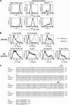
(A) Restoration of the surface expressions of CD59 and DAF after transfection of a PGAP2 cDNA. Mutant Clone 84 cells were transfected with a mock vector or PGAP2 cDNA expression vector. At 2 d after the transfection, the cells were stained with biotinylated anti-CD59 (top) and anti-DAF (bottom) antibodies followed by phycoerythrin-conjugated streptavidin. The dotted lines show negative control staining with the second reagent only. (B) Selective decrease in surface GPI-APs on the mutant cells. Mutant AM-B cells were cotransfected with the indicated plasmids and either a mock vector (bold lines) or PGAP2 expression vector (dotted lines). FLAG-tagged-CD59, FLAG-tagged-folate receptor, and PLAP are GPI-APs, whereas FLAG-tagged-CD59-TM, IL2 receptor α (IL2Rα), p75 (nerve growth factor receptor; NGFR), and FLAG-VSVG are transmembrane (TM) proteins. At 2 d after the transfection, the cells were stained with anti-FLAG, anti-PLAP, anti-IL2Rα, or anti-p75 antibodies. (C) Alignment of the amino acid sequences of PGAP2 homologues. Rat (GenBank Accession No. AB236144), Chinese hamster (Accession No. AB236145), and human (Accession No. AAQ75733) PGAP2s are aligned using the ClustalW software. The 22 nucleotides encoding the amino acids indicated by ♦1 are deleted in the mutant AM-B cells (see Supplementary Information). There is also an alternatively spliced form of PGAP2 that encodes a protein lacking the four amino acids indicated by ♦2.
Figure 3.
Figure 3.
Subcellular localization of PGAP2. (A) NRK cells transfected with the rat PGAP2 cDNA and myc-tagged DPM2 were fixed, permeabilized with 0.1% TX-100, and double-stained with anti-PGAP2 and either anti-GM130 (top) or anti-myc (bottom) antibodies. The bottom panels show staining after treatment with BFA for 10 min. (B) PGAP2 was expressed in AM-B cells at the lowest concentration required for restoration of surface CD59 expression. The cell lysate was layered on top of a continuous sucrose gradient and centrifuged at 35,000 rpm at 4°C for 19 h. Fractions were collected and analyzed by SDS-PAGE and Western blotting.
Figure 4.
Figure 4.
Pulse-chase metabolic labeling of GPI-APs. AM-B and BTP2 cells were pulsed with [35S]methionine and [35S]cysteine for 10 min and then chased for the indicated periods. The cell lysates and culture supernatants were immunoprecipitated with an anti-DAF antibody, separated by SDS-PAGE, and analyzed using a BAS 1000 analyzer.
Figure 5.
Figure 5.
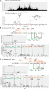
Secretion of GPI-APs by cleavage between inositol and phosphate. (A) HFGF-CD59 secreted from AM-B cells was digested with trypsin and analyzed by LC/ESIMS/MS. Top, the total ion chromatogram; bottom, a mass chromatogram of the molecular ions that generated a GPI-specific fragment ion of m/z 447+ in the MS/MS analysis. The molecular ions labeled 1–4 (peaks 1–4) correspond to C-terminal peptides bearing GPI. (B) MS/MS spectrum of the peak 1 ion of m/z 1226.72+ and its determined structure. Top, the determined structure and the absolute mass; middle, first MS analysis of peak 1; bottom, the second MS of the parent fragment with m/z 1226.72+ shown in the middle panel. The b-series fragments are indicated by orange arrows, and the terminal and internal fragments of GPI structure are indicated by green and gray arrows, respectively. The peak of 1032.9+ in the middle panel corresponded to an internal peptide, LTQS-MAIIR, from HFGF-CD59. (C) MS/MS spectrum of the peak 3 ion of m/z 1328.02+ and its determined structure. Panels are indicated similarly to B. The size difference of m/z between peaks 1 and 3 (101.32+) corresponded to N-acetylhexosamine (HexNAc) attached to the first mannose. Note that the parent fragments with m/z 1032.9+ and 1226.72+ were also present in peak 3 (middle) because of insufficient separation of peak 3 from very close peak 1 in LC (A).
Figure 6.
Figure 6.
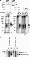
Release of DAF from the membrane at the cell surface. (A) AM-B cells were pulse-labeled with [35S]methionine and [35S]cysteine and chased under the four conditions shown in the top panel. A TX-114 partitioning assay was used to separate the soluble and membrane-bound proteins into aqueous and detergent phases, respectively. The chases under conditions that inhibit transport to the cell surface, such as in the presence of BFA (conditions 1 and 4) or incubation at 19.5°C (conditions 3 and 4), prevent the release of DAF, contrary to the normal condition (condition 2). Cell, cell lysate; M, medium; A, aqueous phase; D, detergent phase; a, premature form; b, secreted form; c, mature form. (B) After pulse-labeling with [35S]methionine and [35S]cysteine for 10 min, AM-B cells were chased with or without 0.3% tannic acid for 40 min. The cell lysates were phase-separated by TX-114 partitioning. C, cell lysate; M, medium; A, aqueous phase; D, detergent phase.
Figure 7.
Figure 7.
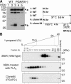
Intracellular processing of a reporter GPI-AP. (A) Wild-type 3B2A (lanes 1–3) and C84 (lanes 4 and 5) cells were transfected without (lane 1) or with (lanes 2–5) VSVGts-FF-mEGFP-GPI and cultured at 40°C for 1 d. Cells were further cultured under the conditions shown on the right. The cell lysates were immunoprecipitated with an anti-FLAG antibody, subjected to SDS-PAGE, and analyzed by Western blotting. a, VSVGts-FF-mEGFP-GPI; b, FLAG-mEGFP-GPI (a cleavage product of VSVGts-FF-mEGFP-GPI by furin); asterisk, a degradation product. (B) 3B2A and C84 cells transfected with VSVGts-FF-mEGFP-GPI were cultured under the same conditions shown in lanes 3 and 5 in A. FLAG-mEGFP-GPI was collected from the cell lysates with anti-FLAG beads and eluted with a FLAG-peptide. To prepare lyso-GPI-anchored FLAG-mEGFP-GPI, aliquots of the immunoprecipitates from 3B2A cells were treated with PLA2. FLAG-mEGFP-GPIs were chromatographed in an Octyl-FF column with a 5–40% gradient of 1-propanol. Fractions were subjected to SDS-PAGE and analyzed by Western blotting with an anti-FLAG antibody.

Similar articles

Cited by

References

    1. Ahmed, S. N., Brown, D. A., and London, E. (1997). On the origin of sphingolipid/cholesterol-rich detergent-insoluble cell membranes: physiological concentrations of cholesterol and sphingolipid induce formation of a detergent-insoluble, liquid-ordered lipid phase in model membranes. Biochemistry 36, 10944–10953. - PubMed
    1. Brewis, I. A., Ferguson, M. A., Mehlert, A., Turner, A. J., and Hooper, N. M. (1995). Structures of the glycosyl-phosphatidylinositol anchors of porcine and human renal membrane dipeptidase. Comprehensive structural studies on the porcine anchor and interspecies comparison of the glycan core structures. J. Biol. Chem. 270, 22946–22956. - PubMed
    1. Brown, D. A., and Rose, J. K. (1992). Sorting of GPI-anchored proteins to glycolipid-enriched membrane subdomains during transport to the apical cell surface. Cell 68, 533–544. - PubMed
    1. Chatterjee, S., Smith, E. R., Hanada, K., Stevens, V. L., and Mayor, S. (2001). GPI anchoring leads to sphingolipid-dependent retention of endocytosed proteins in the recycling endosomal compartment. EMBO J. 20, 1583–1592. - PMC - PubMed
    1. Cheong, K. H., Zacchetti, D., Schneeberger, E. E., and Simons, K. (1999). VIP17/MAL, a lipid raft-associated protein, is involved in apical transport in MDCK cells. Proc. Natl. Acad. Sci. USA 96, 6241–6248. - PMC - PubMed

Publication types

MeSH terms