. 2006 Mar;17(3):1051-64.
doi: 10.1091/mbc.e05-09-0886. Epub 2006 Jan 11.

Conditional dominant mutations in the Caenorhabditis elegans gene act-2 identify cytoplasmic and muscle roles for a redundant actin isoform

Affiliations

Conditional dominant mutations in the Caenorhabditis elegans gene act-2 identify cytoplasmic and muscle roles for a redundant actin isoform

John H Willis et al. Mol Biol Cell. 2006 Mar.

Abstract

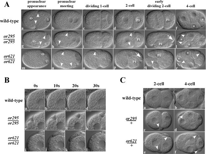
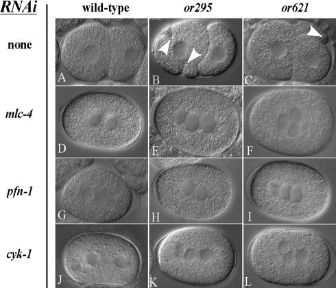
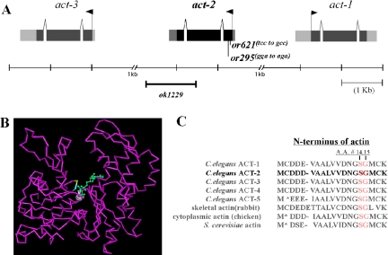
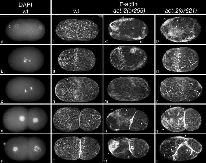
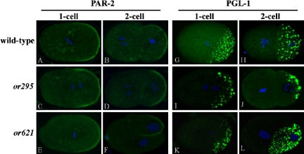
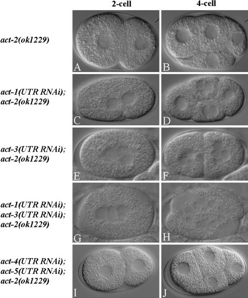
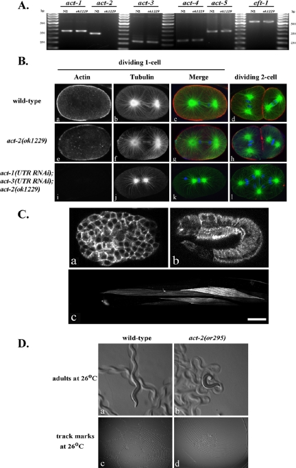
Animal genomes each encode multiple highly conserved actin isoforms that polymerize to form the microfilament cytoskeleton. Previous studies of vertebrates and invertebrates have shown that many actin isoforms are restricted to either nonmuscle (cytoplasmic) functions, or to myofibril force generation in muscle cells. We have identified two temperature-sensitive and semidominant embryonic-lethal Caenorhabditis elegans mutants, each with a single mis-sense mutation in act-2, one of five C. elegans genes that encode actin isoforms. These mutations alter conserved and adjacent amino acids predicted to form part of the ATP binding pocket of actin. At the restrictive temperature, both mutations resulted in aberrant distributions of cortical microfilaments associated with abnormal and striking membrane ingressions and protrusions. In contrast to the defects caused by these dominant mis-sense mutations, an act-2 deletion did not result in early embryonic cell division defects, suggesting that additional and redundant actin isoforms are involved. Accordingly, we found that two additional actin isoforms, act-1 and act-3, were required redundantly with act-2 for cytoplasmic function in early embryonic cells. The act-1 and -3 genes also have been implicated previously in muscle function. We found that an ACT-2::GFP reporter was expressed cytoplasmically in embryonic cells and also was incorporated into contractile filaments in adult muscle cells. Furthermore, one of the dominant act-2 mutations resulted in uncoordinated adult movement. We conclude that redundant C. elegans actin isoforms function in both muscle and nonmuscle contractile processes.


Figures

Figure 1.
Figure 1.
Ectopic membrane ingressions and protrusions in early or295 and or621 mutant embryos. (A) DIC micrographs from time-lapse images of live wild-type (a–f), or295 (g–l) and or621 (m–r) embryos. Arrowheads indicate the pseudocleavage furrow in a wild-type embryo, and abnormal membrane ingressions and protrusions in mutant embryos (a, g, h, j, l, n, o, q, and r). Oocyte (o) and sperm (s) pronuclei in 1-cell embryos are labeled; the posterior daughter of the first mitotic division (P1) is labeled, and the two daughters of P1 are indicated with asterisks. Arrows indicate the direction of cytoplasm protrusion in AB cells of or295 and or621 mutant embryos (k and p). Beginning at the 2-cell stage, these protrusions result in the mis-positioning of embryonic cells in or295 and or621 embryos. In some or621 panels, not all nuclei are present in the focal plane shown (m). In this and all subsequent figures, anterior is to the left and posterior to the right. C. elegans embryos are ∼50 μm in length. (B) Higher magnification DIC photomicrographs of the anterior cortex of the embryonic cell AB in live 2-cell stage wild-type, or295, and or621 embryos. The membrane ingressions in mutant embryos often move along the surface of embryonic cells (or295; e–h), and the protrusions often grow in size over time (or621; i–l). Images were taken every 10 s from time-lapse video micrographs, beginning roughly at the completion of cytokinesis after the first mitotic division of each embryo. (C) Two- and 4-cell stage embryos produced by heterozygous or295/+ and or621/+ mothers raised at the restrictive temperature of 26°C often exhibit abnormal membrane ingressions and protrusions (arrowheads), due to the dominant nature of these mutations (see text for details; c-d and e-f).
Figure 2.
Figure 2.
Ectopic membrane ingressions and protrusions in dominant act-2 mutant embryos require a functional microfilament cytoskeleton. DIC photomicrographs of 2-cell stage wild-type, or295, and or621 embryos (A–C) and equivalent stage wild-type, or295 and or621 embryos after RNAi-mediated depletion of actomyosin function (D–L). Arrowheads indicate ectopic ingressions (B) and a protrusion (C) in the mutant embryos. Depletion of actomyosin function prevents cytokinesis in all embryos and eliminates, or nearly eliminates, abnormal membrane ingressions and protrusions in the mutant embryos (C–F). The presence of more than two nuclei in many of the RNAi-depleted embryos is due to failures of meiotic cytokinesis and consequent aneuploidy (unpublished data). Also see Supplementary Movies 8–13.
Figure 3.
Figure 3.
or295 and or621 are act-2 alleles. (A) The actin gene cluster on linkage group V, where three of the five C. elegans actin genes reside, and the approximate location of the act-2 (ok1229) deletion and the act-2(or295) and act-2(or621) molecular lesions. (B) The act-2(or295) and act-2(or621) mutations alter conserved amino acids in a predicted loop that forms part of the ATP binding pocket of subdomain 1 of actin. The affected residues are highlighted in blue (S14A) and yellow (G15R) and are near the ATP molecule (green), which resides close to the center of the actin structure (purple). The structure, PDB name 1D4X, is from C. elegans Mg-ATP actin, purified from extracts and, thus, not isoform-specific (Vorobiev et al., 2003; see Materials and Methods). (C) The N-terminal amino acid sequences (amino acids 1 through 19 or 20) of different actin isoforms in C. elegans and other eukaryotes. The residues in ACT-2 altered by the or295 (G15R) and or621 (S14A) mutations are highlighted in red. As is conventional for actin, numbers are assigned to amino acid residues after removal of the first two residues (methionine and cysteine), which occurs during posttranslational processing (adapted from Ono, 1999).
Figure 4.
Figure 4.
Cortical microfilaments during the first two embryonic cell cycles in fixed wild-type and dominant act-2 mutant embryos. Wild-type embryos (a–j) costained with DAPI (a–e) to label DNA and Bodipy-fl-phallicidin (f–j) to label microfilaments. act-2(or295) embryos (k–o) and act-2(or621) embryos (p–t) also were stained with Bodipy-fl-phallicidin and DAPI. Embryos in each row are at the same point in the cell cycle, based on DAPI staining of chromosomes (not presented here for the mutant embryos). Each phallicidin-stained image is a maximum projection of 25 confocal sections taken at 0.25-μm intervals. All embryos were fixed and stained using the same procedures, imaged using the same confocal settings, and digitally processed identically. DAPI-stained images are maximum projections of 10–15 widefield epifluorescence images taken at 0.5-μm intervals. Both or295 and or621 embryos contain abnormally dense accumulations of cortical microfilaments (white arrowheads in k, n, o, p, s, and t). These are most pronounced within, but not restricted to, interphase embryos. Regions of dense accumulation often lie adjacent to regions that appear to be depleted of filaments relative to the wild type (white asterisks in k, n, o, p, s, and t), suggesting that microfilaments are abnormally distributed within the cortex. act-2(or621) embryos also accumulate dense microfilaments-containing structures (arrow in q), reminiscent of the barlike structures observed in yeast cells bearing the same point mutation (Chen and Rubenstein, 1995). These structures were mostly present deeper in the cytoplasm (unpublished data), but were also detected at or near the cortical surface. Late in anaphase, cortical microfilaments are reduced in density throughout the cortex outside the cleavage furrow, in both wild-type and mutant embryos (h, m, and r). The accumulation of microfilaments into a contractile ring late in anaphase was not obviously affected in the mutant embryos, although it is not apparent in this example of an or295 mutant embryo (m).
Figure 5.
Figure 5.
Anterior-posterior polarity appears normal in act-2 mutant embryos. Fixed wild-type, act-2(or295), and act-2(or621) embryos stained with fluorescently labeled antibodies that recognize the polarity regulatory protein PAR-2 (A–F) and the P granule component PGL-1 (G–L). Both proteins are localized normally in 1- and 2-cell stage act-2(or295) and act-2(or621) mutant embryos. Note that in F, even though the P1 cell is becoming abnormally positioned, PAR-2 maintains a posterior cortical localization.
Figure 6.
Figure 6.
act-1, -2, and -3 are redundantly required for early embryonic cell divisions. DIC photomicrographs of live 2- and 4-cell stage embryos from mothers homozygous for the act-2(ok1229) deletion (A and B) and equivalent stage embryos from act-2(ok1229) mothers after RNAi-mediated depletion of other actin isoforms, using 3′UTR dsRNA sequences specific for each isoform (see text and Materials and Methods for details). Early cell divisions appeared normal in embryos from act-2(ok1229) worms, although some embryonic lethality occurred in embryos produced at 26°C (Table 1). Depletion of either ACT-1 or -3 in the act-2(ok1229) background resulted in 100% embryonic lethality (Table 2) and in cytokinesis defects and thus multinucleate embryonic cells (C–F). Depletion of both ACT-1 and -3 in act-2(ok1229) embryos resulted in more severe cytokinesis defects (H). Depletion of both ACT-4 and -5 in act-2(ok1229) worms did not result in early cell division defects (I and J). See Table 2 for the number of embryonic cell divisions scored for cytokinesis defects. Also see Supplementary Movies 14–16.
Figure 7.
Figure 7.
Actin gene expression in oocytes, early embryos, and adult muscle; motility defects in act-2(or295) adults. (A) Amplification of actin gene transcripts from isolated oocytes. Nested RT-PCR was performed using gene-specific 3′UTR primer sequences (see Materials and Methods). (B) Immunofluorescence photomicrographs of fixed wild-type and mutant embryos stained with fluorescently labeled antibodies that recognize actin (red) or tubulin (green); DNA (blue) was labeled with TOTO3. Confocal laser strength and antibody concentration were identical in all cases (see Materials and Methods). Little or no actin was detected in act-1(3UTR RNAi); act-3(3UTR RNAi); act-2(ok1229) embryos (i, k, and l). Extra chromosomes in panel k presumably are due to a failure in polar body extrusion during meiosis. (C) Cytoplasmic GFP::ACT-2 expression in live embryos was detected throughout the cytoplasm and at the cortex of all early embryonic cells (a), beginning at about the 20-cell stage (unpublished data), and in epidermal cells during embryonic elongation (b). GFP::ACT-2 also was detected in contractile filaments of adult striated muscle cells (c). Scale bar, 25 μm. (D) Motility defects in act-2(or295) adult animals at the restrictive temperature. Animals were placed on individual plates for 6 h before recording images, and track marks were recorded at 400× and 125× magnification, respectively. Also see Supplementary Movies 14 and 15, in which adult worm movements were recorded at 1 frame/s. Motility defects are apparent for act-2(or295), whereas act-2(or621) mutants resemble wild-type adults in their motility.

Similar articles

Cited by

References

    1. Albertson, D. G. (1984). Formation of the first cleavage spindle in nematode embryos. Dev. Biol. 101, 61–72. - PubMed
    1. Alder, M. N., Dames, S., Gaudet, J., and Mango, S. E. (2003). Gene silencing in Caenorhabditis elegans by transitive RNA interference. RNA 9, 25–32. - PMC - PubMed
    1. Ausubel, F. M., Brent, R., Kingston, R. E., Moore, D. D., Seidman, J. G., Smith, J. A., and Struhl, K. (1991). Current Protocols in Molecular Biology, New York: Greene Publishing Associates and Wiley-Interscience.
    1. Avery, L. (1993). The genetics of feeding in Caenorhabditis elegans. Genetics 133, 897–917. - PMC - PubMed
    1. Bowerman, B., Eaton, B. A., and Priess, J. R. (1992). skn-1, a maternally expressed gene required to specify the fate of ventral blastomeres in the early C. elegans embryo. Cell 68, 1061–1075. - PubMed

Publication types

MeSH terms

LinkOut - more resources