

Musculoskeletal plasticity after acute spinal cord injury: effects of long-term neuromuscular electrical stimulation training
- PMID: 16407424
- PMCID: PMC3298883
- DOI: 10.1152/jn.01181.2005
Musculoskeletal plasticity after acute spinal cord injury: effects of long-term neuromuscular electrical stimulation training
Abstract
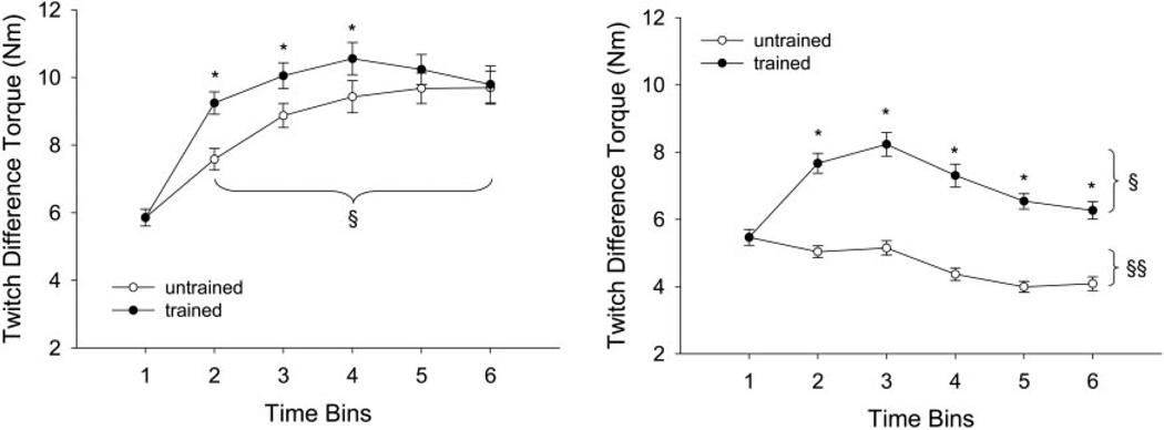
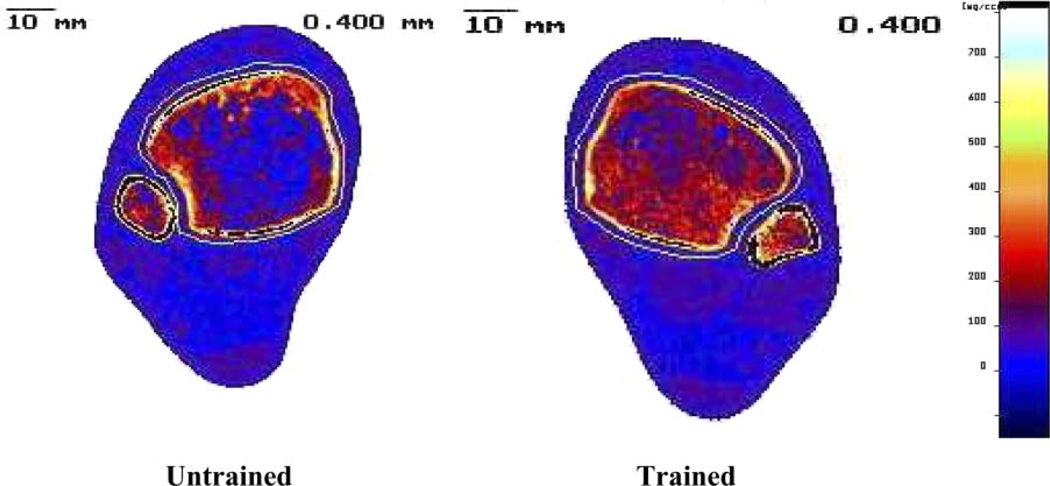
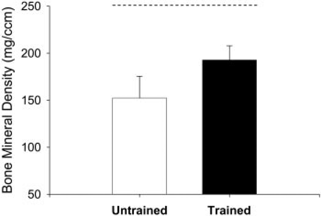
Maintaining the physiologic integrity of paralyzed limbs may be critical for those with spinal cord injury (SCI) to be viable candidates for a future cure. No long-term intervention has been tested to attempt to prevent the severe musculoskeletal deterioration that occurs after SCI. The purposes of this study were to determine whether a long-term neuromuscular electrical stimulation training program can preserve the physiological properties of the plantar flexor muscles (peak torque, fatigue index, torque-time integral, and contractile speed) as well as influence distal tibia trabecular bone mineral density (BMD). Subjects began unilateral plantar flexion electrical stimulation training within 6 wk after SCI while the untrained leg served as a control. Mean compliance for the 2-yr training program was 83%. Mean estimated compressive loads delivered to the tibia were approximately 1-1.5 times body weight. The training protocol yielded significant trained versus untrained limb differences for torque (+24%), torque-time integral (+27%), fatigue index (+50%), torque rise time (+45%), and between-twitch fusion (+15%). These between-limb differences were even greater when measured at the end of a repetitive stimulation protocol (125 contractions). Peripheral quantitative computed tomography revealed 31% higher distal tibia trabecular BMD in trained limbs than in untrained limbs. The intervention used in this study was sufficient to limit many of the deleterious muscular and skeletal adaptations that normally occur after SCI. Importantly, this method of load delivery was feasible and may serve as the basis for an intervention to preserve the musculoskeletal properties of individuals with SCI.
Figures

Similar articles
-
Musculoskeletal adaptations in chronic spinal cord injury: effects of long-term soleus electrical stimulation training.Neurorehabil Neural Repair. 2007 Mar-Apr;21(2):169-79. doi: 10.1177/1545968306293447. Neurorehabil Neural Repair. 2007. PMID: 17312092 Free PMC article.
-
Postfatigue potentiation of the paralyzed soleus muscle: evidence for adaptation with long-term electrical stimulation training.J Appl Physiol (1985). 2006 Aug;101(2):556-65. doi: 10.1152/japplphysiol.00099.2006. Epub 2006 Mar 30. J Appl Physiol (1985). 2006. PMID: 16575026 Free PMC article. Clinical Trial.
-
High dose compressive loads attenuate bone mineral loss in humans with spinal cord injury.Osteoporos Int. 2012 Sep;23(9):2335-46. doi: 10.1007/s00198-011-1879-4. Epub 2011 Dec 21. Osteoporos Int. 2012. PMID: 22187008 Free PMC article.
-
Muscle and bone plasticity after spinal cord injury: review of adaptations to disuse and to electrical muscle stimulation.J Rehabil Res Dev. 2008;45(2):283-96. doi: 10.1682/jrrd.2007.02.0031. J Rehabil Res Dev. 2008. PMID: 18566946 Free PMC article. Review.
-
Muscular, skeletal, and neural adaptations following spinal cord injury.J Orthop Sports Phys Ther. 2002 Feb;32(2):65-74. doi: 10.2519/jospt.2002.32.2.65. J Orthop Sports Phys Ther. 2002. PMID: 11838582 Free PMC article. Review.
Cited by
-
Regional cortical and trabecular bone loss after spinal cord injury.J Rehabil Res Dev. 2012;49(9):1365-76. doi: 10.1682/jrrd.2011.12.0245. J Rehabil Res Dev. 2012. PMID: 23408218 Free PMC article.
-
Effects of Muscles on Bone Metabolism-with a Focus on Myokines.Ann Geriatr Med Res. 2022 Jun;26(2):63-71. doi: 10.4235/agmr.22.0054. Epub 2022 Jun 20. Ann Geriatr Med Res. 2022. PMID: 35722780 Free PMC article.
-
Effects of chronic electrical stimulation on paralyzed expiratory muscles.J Appl Physiol (1985). 2008 Jun;104(6):1634-40. doi: 10.1152/japplphysiol.01321.2007. Epub 2008 Apr 10. J Appl Physiol (1985). 2008. PMID: 18403449 Free PMC article.
-
MicroRNA-208b progressively declines after spinal cord injury in humans and is inversely related to myostatin expression.Physiol Rep. 2015 Nov;3(11):e12622. doi: 10.14814/phy2.12622. Epub 2015 Nov 24. Physiol Rep. 2015. PMID: 26603456 Free PMC article.
-
Altered mRNA expression after long-term soleus electrical stimulation training in humans with paralysis.Muscle Nerve. 2011 Jan;43(1):65-75. doi: 10.1002/mus.21831. Muscle Nerve. 2011. PMID: 21171097 Free PMC article.
References
-
- Aagaard P, Simonsen EB, Andersen JL, Magnusson P, Dyhre-Poulsen P. Neural adaptation to resistance training: changes in evoked V-wave and H-reflex responses. J Appl Physiol. 2002;92:2309–2318. - PubMed
-
- American Spinal Injury Association. International Standards for Neurological Classification of SCI. Atlanta, GA: American Spinal Injury Association; 2002.
-
- Andersen JL, Mohr T, Biering-Sorensen F, Galbo H, Kjaer M. Myosin heavy chain isoform transformation in single fibres from m. vastus lateralis in spinal cord injured individuals: effects of long-term functional electrical stimulation (FES) Pflugers Arch Eur J Physiol. 1996;431:513–518. - PubMed
-
- Belanger M, Stein RB, Wheeler GD, Gordon T, Leduc B. Electrical stimulation: can it increase muscle strength and reverse osteopenia in spinal cord injured individuals? Arch Phys Med Rehabil. 2000;81:1090–1098. - PubMed
-
- Bickel CS, Slade JM, VanHiel LR, Gordon WL, Dudley GA. Variable-frequency-train stimulation of skeletal muscle after spinal cord injury. J Rehabil Res Dev. 2004;41:33–40. - PubMed
Publication types
MeSH terms
Grants and funding
LinkOut - more resources
Full Text Sources
Other Literature Sources
Medical