. 2006 Feb;140(2):512-27.
doi: 10.1104/pp.105.074294. Epub 2006 Jan 11.

The fertilization-induced DNA replication factor MCM6 of maize shuttles between cytoplasm and nucleus, and is essential for plant growth and development

Affiliations

The fertilization-induced DNA replication factor MCM6 of maize shuttles between cytoplasm and nucleus, and is essential for plant growth and development

Thomas Dresselhaus et al. Plant Physiol. 2006 Feb.

Abstract

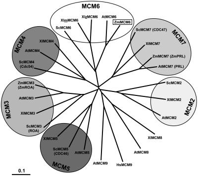
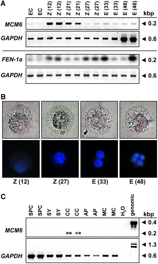
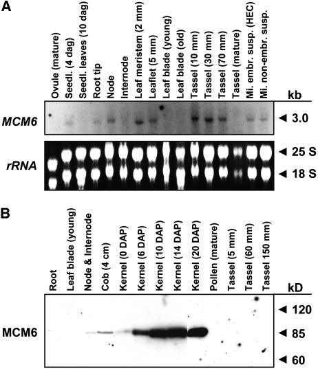
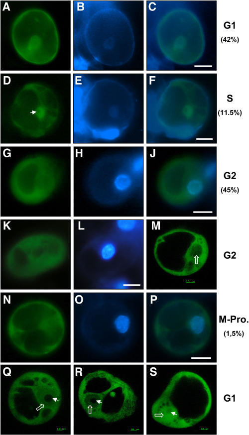
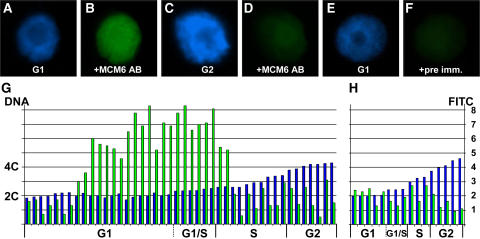
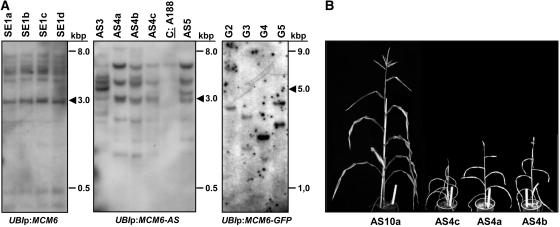
The eukaryotic genome is duplicated exactly once per cell division cycle. A strategy that limits every replication origin to a single initiation event is tightly regulated by a multiprotein complex, which involves at least 20 protein factors. A key player in this regulation is the evolutionary conserved hexameric MCM2-7 complex. From maize (Zea mays) zygotes, we have cloned MCM6 and characterized this essential gene in more detail. Shortly after fertilization, expression of ZmMCM6 is strongly induced. During progression of zygote and proembryo development, ZmMCM6 transcript amounts decrease and are low in vegetative tissues, where expression is restricted to tissues containing proliferating cells. The highest protein amounts are detectable about 6 to 20 d after fertilization in developing kernels. Subcellular localization studies revealed that MCM6 protein shuttles between cytoplasm and nucleoplasm in a cell cycle-dependent manner. ZmMCM6 is taken up by the nucleus during G1 phase and the highest protein levels were observed during late G1/S phase. ZmMCM6 is excluded from the nucleus during late S, G2, and mitosis. Transgenic maize was generated to overexpress and down-regulate ZmMCM6. Plants displaying minor antisense transcript amounts were reduced in size and did not develop cobs to maturity. Down-regulation of ZmMCM6 gene activity seems also to affect pollen development because antisense transgenes could not be propagated via pollen to wild-type plants. In summary, the transgenic data indicate that MCM6 is essential for both vegetative as well as reproductive growth and development in plants.


Figures

Figure 1.
Figure 1.
Phylogenetic tree of MCM2 to 7 protein sequences. Branch lengths are proportional to phylogenetic distances and scale bar represents 10% substitutions per site. The tree was drawn by TreeView from a ClustalW alignment using all available MCM protein sequences from Arabidopsis (AtMCM2-9), maize (ZmMCM3, 6, and 7), frog (XlMCM2-8), budding yeast (ScMCM2-7), as well as human MCM9. Due to historical reasons, some proteins were given two names (old names in parentheses). Note that a maternal (m) and a zygotic (z) MCM6 protein have been described in frog. MCM6 of maize is described in this study. For GenBank accession numbers of sequences, see Table I.
Figure 2.
Figure 2.
ZmMCM6 represents a member of the highly conserved MCM protein family. Homology searches were performed with BLASTP and protein sequences were aligned with ClustalW and drawn using GeneDoc. Identical residues are shaded in black and similar residues are shaded in gray. The alignment shows deduced amino acid sequences of MCM6 from maize (ZmMCM6) and all other eukaryotes available in public databases with the exception of MCM6 from rat, which is almost identical with MCM6 from humans and mouse. GenBank accession numbers for most MCM6 proteins are given in Table I. The human (HsMCM6), mouse (MmMCM6), C. elegans (CeMCM6), and fission yeast (SpMCM6) protein sequence accessions are Q14566, P97311, P34647, and P49731, respectively. Amino acid residues identical/similar in MCM proteins listed in Table I are labeled with asterisks. Highest conserved regions consist of the P-loop, the Walker B motif, and the R- or SRF-finger. The first two motifs are characteristic for ATPases and are essential for NTP binding. The Arg residue within the R-finger probably represents the catalytic activity. The four triangles mark the Cys residues of the zinc-finger-type motif. A predicted cyclin/CDK phosphorylation site that occurs only in plant MCM6 proteins is encircled. A conserved C-terminal region characterized by an aliphatic/polar region of 10 amino acids (dots) flanked on both sides by one to two acidic amino acid residues, which is characteristic for MCM6 of multicellular organisms, but absent in maternal MCM6 of frog and MCM6 proteins of yeast, is boxed with broken lines. The C-terminal region of ZmMCM6 that was used to obtain a peptide antibody is indicated by a horizontal bar.
Figure 3.
Figure 3.
Expression of ZmMCM6 in gametic cells before and after fertilization. A, SC RT-PCR of ZmMCM6 and ZmFEN-1a in egg cells (EC), zygotes (Z), and proembryos (E) at different time points after IVP. The numbers in parentheses indicate hours after pollination (fertilization occurs about 6 h after IVP; top row). Amplification of GAPDH from the same cells is shown for comparison in the corresponding bottom rows. Note that the cells for ZmMCM6 and for DAPI staining (B) were collected in winter and for ZmFEN-1a in summer. ZmFEN1a signals were intensified after blotting and hybridization with a gene-specific radioactive probe. B, Light (top row) and fluorescent images (bottom row) of DAPI-stained zygotes and proembryos. Time points after IVP are indicated. Cell size is about 60 μm. C, SC RT-PCR of ZmMCM6 in sperm cells (SPC), cells of the female gametophyte, including synergid (SY), central cell (CC), and antipodals (AP), as well as leaf mesophyll cell (MC) and controls (water and genomic). Amplification of GAPDH from the same samples is shown in the bottom row.
Figure 4.
Figure 4.
Expression of ZmMCM6 gene and protein in vegetative and complex reproductive tissues. A, RNA gel blot showing expression of ZmMCM6 in the vegetative and reproductive tissues indicated. Fifteen micrograms of total RNA from each tissue were hybridized to a radioactively labeled probe containing the 3′ untranslated region as well as 896 bp encoding the C-terminal region of the gene. The film was exposed for 2 weeks using intensifier screens. B, Protein gel blot incubated with a peptide antibody directed against 15 amino acid residues within a ZmMCM6 C-terminal-specific region (see also Fig. 2). Ten micrograms of protein of the tissues indicated were each separated by 8% SDS-PAGE and blotted onto a polyvinylidene difluoride membrane. A single band was obtained showing the specificity of the antibody.
Figure 5.
Figure 5.
ZmMCM6 protein is excluded from the nucleus in epidermal onion cells. Onion cells were transiently transformed with a UBIp:ZmMCM6-GFP construct and analyzed using epifluorescence (A and C) and light microscopy (B and D). A, Top view of a nucleus showing accumulation of chimeric protein in cytoplasm around the nucleus. The arrows point toward cytoplasm surrounding the nucleus and a transvacuolar cytoplasmic strand. B, Light microscopic image of A. C, Side view of a nucleus showing accumulation of chimeric protein in cytoplasm around the nucleus (arrows), but not inside the nucleus. D, Light microscopic image of C. E, Epifluorescence of an onion epidermal cell bombarded with a 35Sp:Lc-GFP construct encoding the N-terminal 388 amino acids (including the NLS) of a maize transcriptional regulator of anthocyanin biosynthesis in maize (GenBank accession no. A41388) fused with GFP. Most of the fluorescence was detected within the nucleus (arrow). Accumulation of the chimeric protein in cytoplasm around the nucleus, as in A and C, was never observed. Arrowheads in A to D point toward nucleoli.
Figure 6.
Figure 6.
ZmMCM6 shuttles between nucleus and cytoplasm in maize BMS suspension cells in a cell cycle-dependent manner. Suspension cells were transiently transformed with a UBIp:ZmMCM6-GFP construct and analyzed using epifluorescence microscopy (A–L and N–P) as well as CSLM (M, Q–S). Relative DNA content of cells was determined after DAPI staining. A, About 42% cells showed localization of the chimeric protein in both cytoplasm and nucleoplasm. B, DAPI staining of the cell shown in A to display the nucleus. C, Merged image of A and B showing similar DAPI signal in the nucleus compared to GFP fluorescence. These cells were counted for G1 phase. D, About 11.5% of the cells examined accumulated most of the chimeric protein in the nucleoplasm, but not in the nucleolus (arrowhead). E, DAPI staining of the cell shown in D. The image was enhanced to display the weak fluorescence of the nucleus in late G1/S phase. F, Merged image of D and E showing mainly GFP fluorescence in the nucleus. G and K, Majority of cells (about 45%) displayed GFP fluorescence only in the cytoplasm and lacked GFP signals in the nucleus. H and L, Strong DAPI staining of the nuclei of these cells revealed that they were in G2 phase. J, Merged image of G and H showing GFP fluorescence in the cytoplasm and DAPI fluorescence in the nucleus. M, Stack of five CLSM images of a cell in G2 phase confirms lack of GFP fluorescence in the nucleus (open white arrow). N, Cell showing GFP fluorescence exclusively in the cytoplasm displayed condensation of chromosomes (O) during prophase of mitosis. P, Merged image of N and O confirms lack of GFP fluorescence in the nucleus. Q and R, Gradient of the fusion protein between cytoplasm and nucleoplasm was observed in stacks of five images each of two different cells in G1 phase. The weak DAPI staining of these cells is not shown. Note that the chimeric protein is localized in both cytoplasm and nucleoplasm (open white arrows), but never in the nucleolus (arrowhead). S, Control showing a suspension cell bombarded with a UBIp:GFP construct showing GFP fluorescence in the nucleolus, nucleoplasm, and cytoplasm. The arrowhead points toward the nucleolus and the open arrow toward the nucleoplasm. Scale bars are 20 μm, unless otherwise indicated.
Figure 7.
Figure 7.
ZmMCM6 is taken up by the nucleus during G1 phase and highest protein levels are detectable during late G1/early S phase of the cell cycle. Relative DNA content of isolated BMS nuclei were determined after DAPI staining. ZmMCM6 protein amounts were measured after incubation with an FITC-coupled antibody against a ZmMCM6 peptide antibody. DAPI staining (A) and FITC signals (B) of a nucleus in G1; DAPI staining (C) and FITC signals (D) of a nucleus in G2; DAPI staining of a G1 phase nucleus (E) and the same nucleus after incubation with preimmune serum showing minor background signals (F). G, Summary of measurements of 44 nuclei. After DAPI measurements, nuclei were classified as G1 (2C ± 15%), G1/S (2C + 16%–25%), S (2C + 26%–84%), and as G2 (4C ± 15%). Histograms show relative DNA (blue bar) and FITC (green bar) levels of individual nuclei. Relative DNA content is indicated at the left and relative FITC signal intensity at the right of image H. H, To show antibody specificity and to determine background signals, nuclei have been incubated with preimmune serum. Classification of nuclei was as described in G.
Figure 8.
Figure 8.
Molecular and phenotypical analysis of transgenic maize containing the ZmMCM6 gene under control of the strong and constitutive maize UBI promoter. A, Examples of Southern blots containing 10 μg genomic DNA of transgenic lines with ZmMCM6 constructs in sense (SE, left) and antisense (AS, middle) orientation, or fused with GFP (G, right). Genomic DNA was digested with enzyme combinations to display full-copy integrations (arrowheads). Left and middle blots were hybridized with a ZmMCM6-specific probe and the right blot with a GFP-specific probe. The control (A188) on the blot in the middle shows the endogenous ZmMCM6 signal. B, Transgenic plants containing a functional ZmMCM6 AS construct were small in size and did not develop cobs to maturity. Tassels contained little pollen. Here, the clonal line AS4a to AS4c is shown. The line AS10a lacking a functional transgene is shown at the left. Plant height of AS10a was comparable to wild-type plants (A188; 160–190 cm) and both a cob and tassel were fully developed. The white ruler in flower pots is 25 cm.

Similar articles

Cited by

References

    1. Arias EE, Walter JC (2005) Replication-dependent destruction of Cdt1 limits DNA replication to a single round per cell cycle in Xenopus egg extracts. Genes Dev 19: 114–126 - PMC - PubMed
    1. Bailis JM, Forsburg SL (2004) MCM proteins: DNA damage, mutagenesis and repair. Curr Opin Genet Dev 14: 17–21 - PubMed
    1. Bastida M, Puigdomènech P (2002) Specific expression of ZmPRL, the maize homolog of MCM7, during early embryogenesis. Plant Sci 162: 97–106
    1. Blow JJ, Dutta A (2005) Preventing re-replication of chromosomal DNA. Nat Rev Mol Cell Biol 6: 476–486 - PMC - PubMed
    1. Bogan JA, Natale DA, Depamphilis ML (2000) Initiation of eukaryotic DNA replication: conservative or liberal? J Cell Physiol 184: 139–150 - PubMed

Publication types

MeSH terms

Associated data