Comparative Study
. 2006 Feb;140(2):411-32.
doi: 10.1104/pp.105.073783. Epub 2006 Jan 11.

Genome-wide analysis of the ERF gene family in Arabidopsis and rice

Affiliations
Comparative Study

Genome-wide analysis of the ERF gene family in Arabidopsis and rice

Toshitsugu Nakano et al. Plant Physiol. 2006 Feb.

Abstract

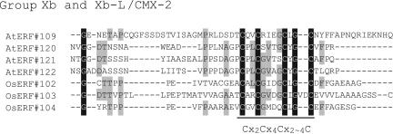
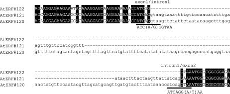
Genes in the ERF family encode transcriptional regulators with a variety of functions involved in the developmental and physiological processes in plants. In this study, a comprehensive computational analysis identified 122 and 139 ERF family genes in Arabidopsis (Arabidopsis thaliana) and rice (Oryza sativa L. subsp. japonica), respectively. A complete overview of this gene family in Arabidopsis is presented, including the gene structures, phylogeny, chromosome locations, and conserved motifs. In addition, a comparative analysis between these genes in Arabidopsis and rice was performed. As a result of these analyses, the ERF families in Arabidopsis and rice were divided into 12 and 15 groups, respectively, and several of these groups were further divided into subgroups. Based on the observation that 11 of these groups were present in both Arabidopsis and rice, it was concluded that the major functional diversification within the ERF family predated the monocot/dicot divergence. In contrast, some groups/subgroups are species specific. We discuss the relationship between the structure and function of the ERF family proteins based on these results and published information. It was further concluded that the expansion of the ERF family in plants might have been due to chromosomal/segmental duplication and tandem duplication, as well as more ancient transposition and homing. These results will be useful for future functional analyses of the ERF family genes.


Figures

Figure 1.
Figure 1.
Alignment of the AP2/ERF domains from Arabidopsis ERF proteins. Black and light gray shading indicate identical and conserved amino acid residues, respectively. Dark gray shading indicates conserved amino acid residues in group VI-L or group Xb-L. The black bar and arrows represent predicted α-helix and β-sheet regions, respectively, within the AP2/ERF domain (Allen et al., 1998). Asterisks represent amino acid residues that directly make contact with DNA (Allen et al., 1998).
Figure 2.
Figure 2.
An unrooted phylogenetic tree of Arabidopsis ERF proteins. The amino acid sequences of the AP2/ERF domain, except members of group VI-L and Xb-L, were aligned by ClustalW (Supplemental Fig. 1), and the phylogenetic tree was constructed using the NJ method. The names of the ERF genes that have already been reported are indicated. The so-called CBF/DREB and ERF subfamilies are divided with a broken line. Classification by Sakuma et al. (2002) is indicated in parentheses.
Figure 3.
Figure 3.
Phylogenetic relationships among the Arabidopsis ERF genes, from group I (A), group II (B), group III (C), group IV (D), group V (E), group VI (F), group VI-L (G), group VII (H), group VIII (I), group IX (J), group X (K), and group Xb-L (L) in the Arabidopsis ERF family. Bootstrap values from 100 replicates were used to assess the robustness of the trees. Bootstrap values >50 are shown. The phylogenetic tree, location of the intron (arrowhead), and a schematic diagram of the protein structures of every group, I to VI, VI to L, VII to X, and Xb-L, are shown in A to L, respectively. Each colored box represents the AP2/ERF domain and conserved motifs, as indicated below the tree. The amino acid sequences of the conserved motifs are summarized in Supplemental Table IV. The asterisk indicates that these motifs were defined by multiple alignments with manual correction rather than an MEME search. Classification by Sakuma et al. (2002) is indicated in parentheses.
Figure 3.
Figure 3.
Phylogenetic relationships among the Arabidopsis ERF genes, from group I (A), group II (B), group III (C), group IV (D), group V (E), group VI (F), group VI-L (G), group VII (H), group VIII (I), group IX (J), group X (K), and group Xb-L (L) in the Arabidopsis ERF family. Bootstrap values from 100 replicates were used to assess the robustness of the trees. Bootstrap values >50 are shown. The phylogenetic tree, location of the intron (arrowhead), and a schematic diagram of the protein structures of every group, I to VI, VI to L, VII to X, and Xb-L, are shown in A to L, respectively. Each colored box represents the AP2/ERF domain and conserved motifs, as indicated below the tree. The amino acid sequences of the conserved motifs are summarized in Supplemental Table IV. The asterisk indicates that these motifs were defined by multiple alignments with manual correction rather than an MEME search. Classification by Sakuma et al. (2002) is indicated in parentheses.
Figure 4.
Figure 4.
The EAR motif-like sequences conserved in the C-terminal region of subgroup VIIIa and subgroup IIa ERF proteins. A, An alignment of the sequences of the C-terminal regions of subgroup VIIIa proteins. B, An alignment of the sequences of the C-terminal regions of subgroup IIA proteins. The conserved motifs are underlined. Black and gray shading indicate identical and conserved amino acid residues present in more than 50% of the aligned sequences, respectively. Consensus amino acid residues are given below the alignment. The “x” in the sequence indicates no conservation at this position. Bold letters in the sequence represent conserved amino acid residues in the original EAR motif (Ohta et al., 2001). Asterisks indicate proteins with demonstrated repression activity (Fujimoto et al., 2000; Ohta et al., 2001).
Figure 5.
Figure 5.
A putative zinc-finger motif conserved in group Xb and group Xb-L proteins. Black and gray shading indicate identical and conserved amino acid residues present in more than 50% of the aligned sequences, respectively. Consensus amino acid residues are given below the alignment. The “x” indicates no conservation at this position.
Figure 6.
Figure 6.
Putative protein kinase phosphorylation sites conserved in ERF proteins. A, Putative MAP kinase and/or casein kinase I phosphorylation sites conserved in group VI and VI-L proteins. B, Putative MAP kinase phosphorylation sites conserved in group VII proteins. C, Putative MAP kinase phosphorylation sites conserved in subgroup IXb proteins. The conserved motifs are underlined. Black and gray shading indicate identical and conserved amino acid residues present in more than 50% of the aligned sequences, respectively. The “+” indicates an amino acid matching the motif patterns provided by the ELM (http://elm.eu.org/browse.html).
Figure 7.
Figure 7.
The nucleotide sequence alignment of the exon1/intron1/exon2 region in group Xb-L genes. Intron sequences are in lowercase. Putative duplication sites are underlined, and conserved nucleotide sequences are shown. Identical nucleotides are highlighted with black shading.
Figure 8.
Figure 8.
An unrooted phylogenetic tree of the ERF proteins in rice. The amino acid sequences of the AP2/ERF domain, except members of group VI-L, were aligned using ClustalW (Supplemental Fig. 2), and the phylogenetic tree was constructed using the NJ method. The names of the ERF genes that have been reported previously are indicated. The so-called CBF/DREB and ERF subfamilies are divided with a broken line.
Figure 9.
Figure 9.
Conserved amino acid sequence motifs in group VII ERF proteins. A, CMVII-6; B, CMVII-7; C, CMVII-8. The conserved motifs are underlined. Consensus sequences calculated by MEME program are given below the underlines. These regions were identified by MEME search using all members of Arabidopsis and rice group VII proteins. Black and gray shading indicate identical and conserved amino acid residues present in more than 50% of the aligned sequences, respectively.
Figure 10.
Figure 10.
The locations of the ERF family genes on the Arabidopsis chromosomes. The chromosomal positions of the ERF genes are indicated by their generic names (Supplemental Table II). Group/subgroup names are shown in parentheses ahead of the generic name. The chromosome number is indicated at the top of each chromosome. The blue boxes indicate the duplicated segmental regions resulting from the most recent polyploidy (Blanc et al., 2003). Only the duplicated regions containing ERF genes are shown. Identical colored circles or squares indicate duplicated gene pairs, deduced by Blanc et al. (2003). The thick lines join tandem repeated genes. Colored fonts indicate ERF genes located on ancient segmental duplications (Blanc et al., 2003). AtERF#077 and #080 (black triangle), AtERF#017 and #018 (red triangle), AtERF#069 and #070 (blue triangle), and AtERF#051 and #052 (green triangle) are potential duplicated gene pairs, as described in the text.

References

    1. Aharoni A, Dixit S, Jetter R, Thoenes E, van Arkel G, Pereira A (2004) The SHINE clade of AP2 domain transcription factors activates wax biosynthesis, alters cuticle properties, and confers drought tolerance when overexpressed in Arabidopsis. Plant Cell 16: 2463–2480 - PMC - PubMed
    1. Allen MD, Yamasaki K, Ohme-Takagi M, Tateno M, Suzuki M (1998) A novel mode of DNA recognition by a beta-sheet revealed by the solution structure of the GCC-box binding domain in complex with DNA. EMBO J 17: 5484–5496 - PMC - PubMed
    1. Alonso JM, Stepanova AN, Leisse TJ, Kim CJ, Chen H, Shinn P, Stevenson DK, Zimmerman J, Barajas P, Cheuk R, et al (2003) Genome-wide insertional mutagenesis of Arabidopsis thaliana. Science 301: 653–657 - PubMed
    1. Altschul SF, Gish W, Miller W, Myers EW, Lipman DJ (1990) Basic local alignment search tool. J Mol Biol 215: 403–410 - PubMed
    1. Altschul SF, Madden TL, Schäffer AA, Zhang J, Zhang Z, Miller W, Lipman DJ (1997) Gapped BLAST and PSI-BLAST: a new generation of protein database search programs. Nucleic Acids Res 25: 3389–3402 - PMC - PubMed

Publication types