

Axonal growth and guidance defects in Frizzled3 knock-out mice: a comparison of diffusion tensor magnetic resonance imaging, neurofilament staining, and genetically directed cell labeling
- PMID: 16407530
- PMCID: PMC6674392
- DOI: 10.1523/JNEUROSCI.3221-05.2006
Axonal growth and guidance defects in Frizzled3 knock-out mice: a comparison of diffusion tensor magnetic resonance imaging, neurofilament staining, and genetically directed cell labeling
Abstract
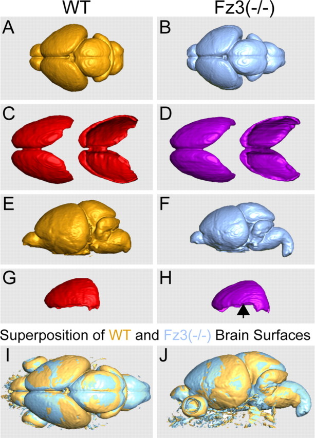
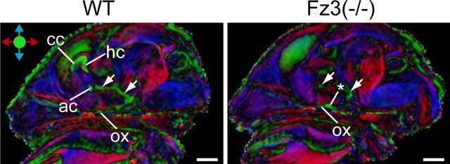
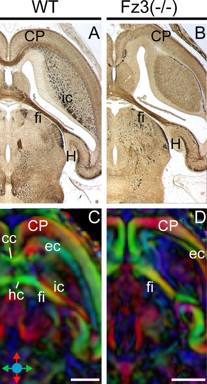
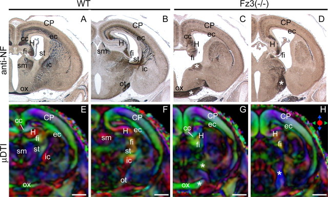
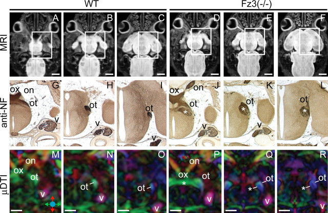
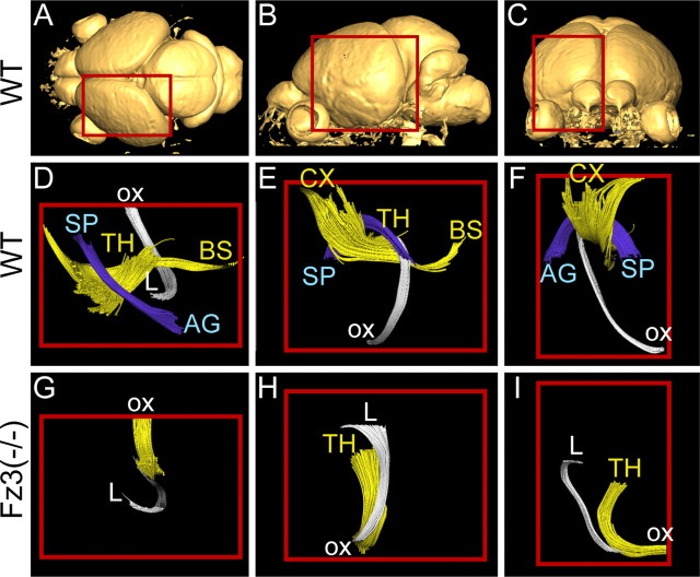
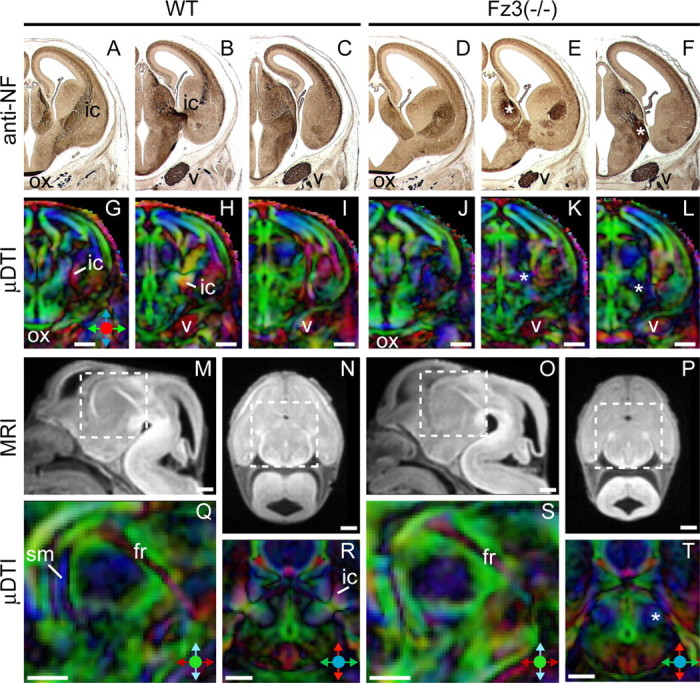
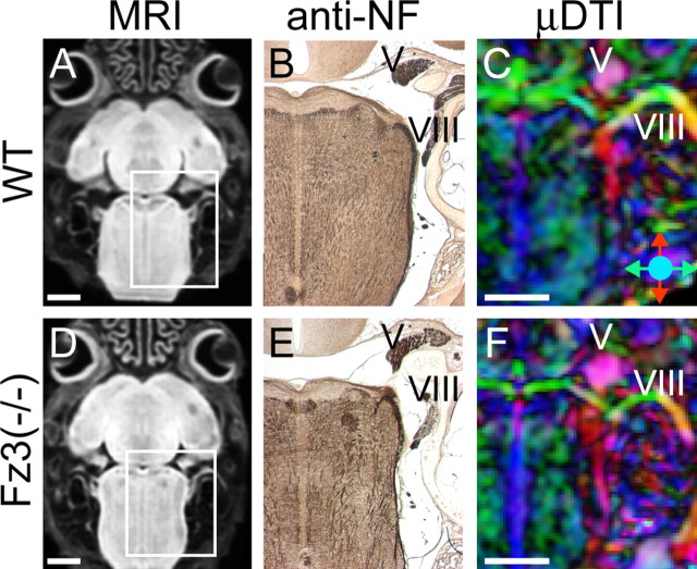
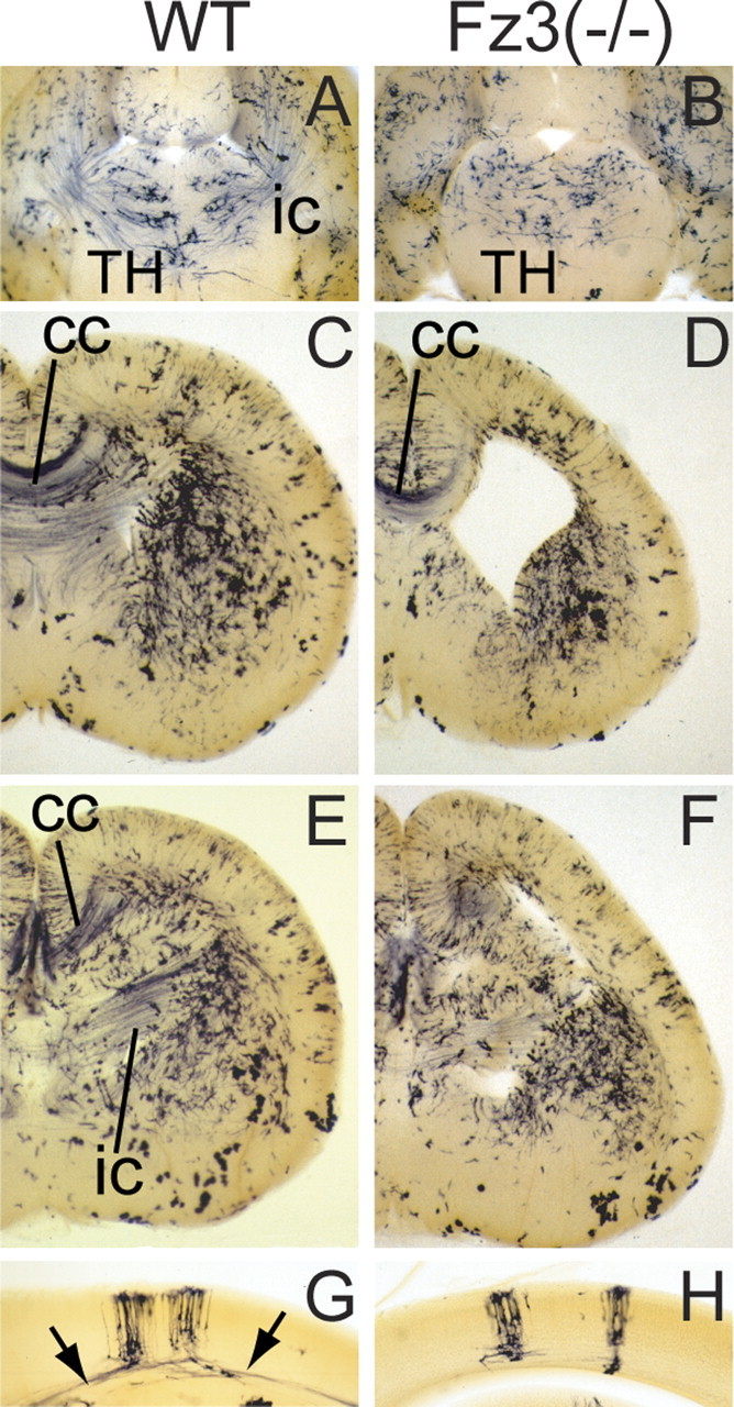
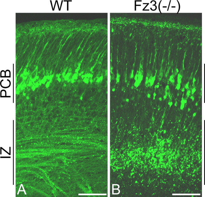
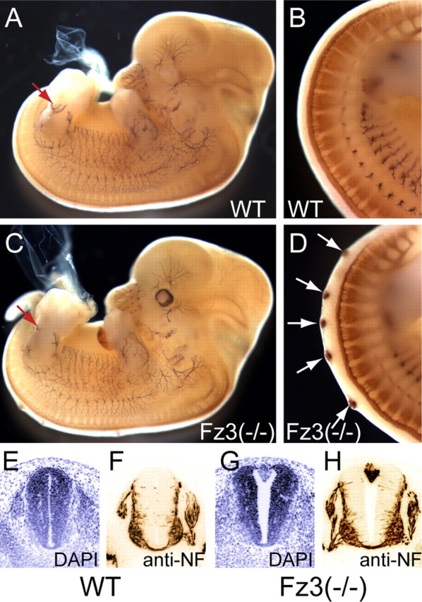
Previous work has identified axonal outgrowth and/or guidance defects in the brain and spinal cord of prenatal Frizzled3 (Fz3)(-/-) mice. To systematically explore the axonal defects in Fz3(-/-) mice and to compare techniques for the global assessment of axon tracts in the developing mouse, we have analyzed wild-type and Fz3(-/-) brains using (1) diffusion tensor magnetic resonance imaging (muDTI), (2) neurofilament staining, and (3) two genetically directed neuronal labeling methods. Confirming and extending the previous work of Wang et al. (2002), we find that the following structures/tracts are absent or greatly reduced in the Fz3(-/-) brain: the anterior commissure, cerebral peduncle (corticospinal tract), corpus callosum, fornix, internal capsule (thalamocortical and corticothalamic tracts), stria medullaris, stria terminalis, and hippocampal commissure. An aberrant U-shaped fiber bundle immediately caudal to the optic tract connects the left and right sides of the Fz3(-/-) thalamus and likely represents a default pathway for thalamic axons that failed to enter the internal capsule. At embryonic day 18, labeling of cortical pyramidal cells with a yellow fluorescent protein reporter reveals widespread fragmentation of axons with no apparent loss of pyramidal cell bodies. Fragmentation likely represents one stage in the process that normally eliminates stalled or mistargeted axons. This work demonstrates the usefulness of muDTI and genetically directed neuronal labeling for the analysis of nervous system defects in the mouse.
Figures
References
-
- Beirowski B, Berek L, Adalbert R, Wagner D, Grumme DS, Addicks K, Ribchester RR, Coleman MP (2004) Quantitative and qualitative analysis of Wallerian degeneration using restricted axonal labelling in YFP-H mice. J Neurosci Methods 134: 23–35. - PubMed
-
- Chae J, Kim MJ, Goo JH, Collier S, Gubb D, Charlton J, Adler PN, Park WJ (1999) The Drosophila tissue polarity gene starry night encodes a member of the protocadherin family. Development 126: 5421–5429. - PubMed
-
- Curtin JA, Quint E, Tsipouri V, Arkell RM, Cattanach B, Copp AJ, Henderson DJ, Spurr N, Stanier P, Fisher EM, Nolan PM, Steel KP, Brown SD, Gray IC, Murdoch JN (2003) Mutation of Celsr1 disrupts planar polarity of inner ear hair cells and causes severe neural tube defects in the mouse. Curr Biol 13: 1129–1133. - PubMed
Publication types
MeSH terms
Substances
Grants and funding
LinkOut - more resources
Full Text Sources
Other Literature Sources
Molecular Biology Databases