. 2006 Jan 11;26(2):389-97.
doi: 10.1523/JNEUROSCI.1203-05.2006.

Late-onset leanness in mice with targeted ablation of melanin concentrating hormone neurons

Affiliations

Late-onset leanness in mice with targeted ablation of melanin concentrating hormone neurons

Tamar Alon et al. J Neurosci. .

Abstract

The observation that loss of orexin (hypocretin) neurons causes human narcolepsy raises the possibility that other acquired disorders might also result from loss of hypothalamic neurons. To test this possibility for body weight, mice with selective loss of melanin concentrating hormone (MCH) neurons were generated. MCH was chosen to test because induced mutations of the MCH gene in mice cause hypophagia and leanness. Mice with ablation of MCH neurons were generated using toxin (ataxin-3)-mediated ablation strategy. The mice appeared normal but, after 7 weeks, developed reduced body weight, body length, fat mass, lean mass, and leptin levels. Leanness was characterized by hypophagia and increased energy expenditure. To study the role of MCH neurons on obesity secondary to leptin deficiency, we generated mice deficient in both ob gene product (leptin) and MCH neurons. Absence of MCH neurons in ob/ob mice improved obesity, diabetes, and hepatic steatosis, suggesting that MCH neurons are important mediators of the response to leptin deficiency. These data show that loss of MCH neurons can lead to an acquired leanness. This has implications for the pathogenesis of acquired changes of body weight and might be considered in clinical settings characterized by substantial weight changes later in life.


Figures
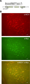
Figure 1.
Figure 1.
Specific expression of ataxin-3 transgene in MCH-containing neurons in MCH/ataxin-3 transgenic mice. A, Schematic of a modified MCH/ataxin-3 BAC construct. A modified MCH BAC contains 76 kb of a 5′ MCH sequence, followed by an ataxin-3 transgene. The transgene contains HA epitope, a cDNA fragment starting at amino acid 286 and ending at the C terminus of human mutant ataxin-3, and MYC epitope followed by a murine protamine 1 gene fragment (mPrm1). B, Double immunohistochemistry showing colocalization of MYC epitope (green fluorescence) and MCH (red fluorescence) in the lateral hypothalamic area (LH) of a 4-week-old MCH/ataxin-3 transgenic mouse. Bottom, Bright yellow indicates the presence of nuclear aggregates stained by anti-MYC antibody with MCH-containing neurons. Scale bar, 100μm.
Figure 2.
Figure 2.
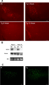
Temporal loss of MCH neurons accompanies the normal expression of orexin in the hypothalamus of MCH/ataxin-3 transgenic mice. A, Immunostaining of matched brain sections from 4-, 8-, and 15-week-old MCH/ataxin-3 transgenic mice (right) and their 4-week-old wild-type littermate (left) using anti-MCH antiserum. Scale bar, 200 μm. B, Northern blot analysis comparing the expression levels of MCH and orexin mRNAs in the hypothalamus of 8- and 36-week-old MCH/ataxin-3 mice and their wild-type littermates. An 18S ribosomal mRNA was used to confirm equal loading. C, Anti-orexin immunostaining of matched brain sections of the lateral hypothalamic area from 8-week-old MCH/ataxin-3 mice (right) and their wild-type littermates (left). Scale bar, 200 μm. Tg, Transgenic; Wt, WT, or W, wild-type.
Figure 3.
Figure 3.
Growth, body composition, and body length in MCH-ataxin-3 mice maintained on a regular diet. A, Weight curves of male MCH/ataxin-3 mice (open squares) and their wild-type littermates (filled diamonds) mice over the course of 23 weeks. n = 6 for each group. *p < 0.005, #p < 0.05. B, Weight curves of female MCH/ataxin-3 mice (open squares) and their wild-type littermates (filled diamonds) mice over the course of 24 weeks. n = 6 for each group. *p < 0.005, #p < 0.05. C, Body composition analysis of both 13-week-old male and 18-week-old female MCH/ataxin-3 (shingle-filled bars and dot-filled bars) and their wild-type littermates (downward diagonal filled bars and solid bars) mice showing fat mass, lean mass, and total water. n = 6 per group, *p < 0.05 versus wild-type control, **p < 0.005. D, Nose–anus length of male and female MCH/ataxin-3 mice (filled bars) and their wild-type littermates (downward diagonal filled bars) at 20 weeks of age (n = 6 in each group). *p < 0.002. Tg, Transgenic; Wt, wild-type.
Figure 4.
Figure 4.
Food intake and metabolic rate in MCH/ataxin-3 transgenic mice. A, Average daily food intake of both male and female MCH/ataxin-3 (shingle-filled bars and dot-filled bars) and their wild-type littermates (downward diagonal filled bars and solid bars) mice at 8, 12, and 16 weeks of age. *p < 0.03 (n = 7). B, Oxygen consumption (in 30 min intervals) of 17-week-old male MCH/ataxin-3 mice (open squares) and their wild-type littermates (filled diamonds) during a 24 h period as measured by an Oxymax monitoring system (n = 6 in each group). Tg, Transgenic; WT, wild-type.
Figure 5.
Figure 5.
Hypothalamic neuropeptide mRNAs in MCH/ataxin-3 mice. Expression levels of POMC, NPY, and AGRP in the hypothalamus of 19-week-old female MCH/ataxin-3 mice (filled bars; n = 6) mice and their wild-type (Wt) littermates (downward diagonal filled bars; n = 6) using a real-time quantitative reverse transcription-PCR. Tg, Transgenic.
Figure 6.
Figure 6.
MCH/ataxin-3 mice exhibited a normal response to fasting, leptin treatment, and leptin deficiency. A, Cumulative food intake in response to 24 h starvation in 19-week-old male MCH/ataxin-3 mice (open squares; n = 5) and their wild-type littermates (wt; filled diamonds; n = 6). *p < 0.03, **p < 0.007. B, C, Food intake (B) and body weight (C) in response to 9 d of leptin treatment followed by 8 d of leptin withdrawal in 19-week-old male MCH/ataxin-3 mice (open squares; n = 6) and their wild-type littermates (filled diamonds; n = 6) compared with PBS control mice (filled triangles; n = 4). Food intake and body weight are represented as a percentage of the baseline. The food intake baseline was an average of the food consumption for 24 h measured over a 3 d period before leptin treatment. The body weight baseline was body weight measured 2 d before leptin treatment. Tg, Transgenic.
Figure 7.
Figure 7.
The loss of MCH neurons in ob/ob mice improved obesity, diabetes, and hepatic abnormalities. A, Body weight in 24- to 28-week-old female MCH/ataxin-3/ob/ob (filled bar; n = 7) and ob/ob control (downward diagonal filled bar; n = 10) mice. *p < 0.004. B, Glucose levels in 24- to 28-week-old female MCH/ataxin-3/ob/ob (filled bar; n = 10), ob/ob control mice (downward diagonal filled bar; n = 8), and wild-type mice (wt; dot-filled bar; n = 6). *p < 0.03, **p < 0.002. C, Photographs of freshly dissected livers from 24-week-old female MCH/ataxin-3/ob/ob (center), ob/ob control (left), and wild-type (right) mice. D, Hematoxylin and eosin staining of liver sections from female MCH/ataxin-3/ob/ob (right) and ob/ob control (left) (females, n = 3; males, n = 2). Scale bar, 20 μm.

Similar articles

Cited by

References

    1. Bevivino AE, Loll PJ (2001) An expanded glutamine repeat destabilizes native ataxin-3 structure and mediates formation of parallel beta-fibrils. Proc Natl Acad Sci USA 98: 11955–11960. - PMC - PubMed
    1. Bittencourt JC, Presse F, Arias C, Peto C, Vaughan J, Nahon JL, Vale W, Sawchenko PE (1992) The melanin-concentrating hormone system of the rat brain: an immuno- and hybridization histochemical characterization. J Comp Neurol 319: 218–245. - PubMed
    1. Bornfeldt KE, Arnqvist HJ, Enberg B, Mathews LS, Norstedt G (1989) Regulation of insulin-like growth factor-I and growth hormone receptor gene expression by diabetes and nutritional state in rat tissues. J Endocrinol 122: 651–656. - PubMed
    1. Borsu L, Presse F, Nahon JL (2000) The AROM gene, spliced mRNAs encoding new DNA/RNA-binding proteins are transcribed from the opposite strand of the melanin-concentrating hormone gene in mammals. J Biol Chem 275: 40576–40587. - PubMed
    1. Bray GA, Gallagher Jr TF (1975) Manifestations of hypothalamic obesity in man: a comprehensive investigation of eight patients and a review of the literature. Med 54: 301–330. - PubMed

Publication types

MeSH terms

LinkOut - more resources