

Expression of a familial Alzheimer's disease-linked presenilin-1 variant enhances perforant pathway lesion-induced neuronal loss in the entorhinal cortex
- PMID: 16407539
- PMCID: PMC6674394
- DOI: 10.1523/JNEUROSCI.3961-05.2006
Expression of a familial Alzheimer's disease-linked presenilin-1 variant enhances perforant pathway lesion-induced neuronal loss in the entorhinal cortex
Abstract
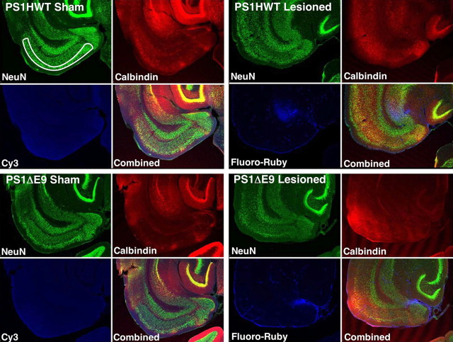
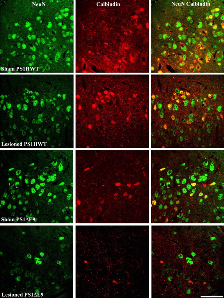
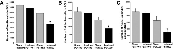
Alzheimer's disease (AD) is characterized by neuronal loss in the hippocampus and entorhinal cortex that is manifested by progressive memory impairment and cognitive decline. Autosomal-dominant, familial forms of AD (FAD) are caused by mutations in genes encoding amyloid precursor protein, presenilin-1 (PS1), and presenilin 2. Although it is established that expression of mutant PS1 variants leads to increased production of highly fibrillogenic amyloidbeta42 (Abeta42) peptides that deposit in the brains of patients with AD, the mechanism(s) by which Abeta deposition and expression of mutant genes induce lamina- and region-specific vulnerability of neuronal populations is not known. We have examined the hypothesis that expression of transgene-encoded FAD-linked mutant PS1 variants in entorhinal cortex neurons exacerbates the vulnerability of these cells to lesion-induced neuronal loss. To test this notion, we transected the perforant pathway (PP) of transgenic mice harboring either wild-type human PS1 (PS1HWT) or the FAD-linked mutant PS1DeltaE9 variant and examined neuronal survival in layer II of the entorhinal cortex (ECL2). Remarkably, PP transections lead to marked reductions in the numbers of ECL2 neurons in the ECL2 of mice expressing mutant PS1, compared with ECL2 neurons in PP-lesioned PS1HWT mice. Finally, and in contrast to studies in nontransgenic mice and in mice expressing PS1HWT, ECL2 neurons that express mutant PS1 and the calcium binding protein calbindin-D28k in ECL2 are also susceptible to lesion-induced neuronal loss. We conclude that expression of FAD-linked mutant PS1 variants enhances the vulnerability of neurons in the entorhinal cortex to PP lesion-induced cytotoxicity.
Figures
Similar articles
-
Fibroblast growth factor-2 protects entorhinal layer II glutamatergic neurons from axotomy-induced death.J Neurosci. 1996 Feb 1;16(3):886-98. doi: 10.1523/JNEUROSCI.16-03-00886.1996. J Neurosci. 1996. PMID: 8558257 Free PMC article.
-
The presenilin-1 familial Alzheimer disease mutant P117L impairs neurogenesis in the hippocampus of adult mice.Exp Neurol. 2004 Aug;188(2):224-37. doi: 10.1016/j.expneurol.2004.04.002. Exp Neurol. 2004. PMID: 15246822
-
Age-related amyloid beta deposition in transgenic mice overexpressing both Alzheimer mutant presenilin 1 and amyloid beta precursor protein Swedish mutant is not associated with global neuronal loss.Am J Pathol. 2000 Jul;157(1):331-9. doi: 10.1016/s0002-9440(10)64544-0. Am J Pathol. 2000. PMID: 10880403 Free PMC article.
-
Metabolism of presenilin 1: influence of presenilin 1 on amyloid precursor protein processing.Neurobiol Aging. 1998 Jan-Feb;19(1 Suppl):S15-8. doi: 10.1016/s0197-4580(98)00026-8. Neurobiol Aging. 1998. PMID: 9562461 Review.
-
Death and survival of neuronal cells exposed to Alzheimer's insults.J Neurosci Res. 2002 Nov 1;70(3):380-91. doi: 10.1002/jnr.10354. J Neurosci Res. 2002. PMID: 12391601 Review.
Cited by
-
A meeting to remember: meeting on memory and related disorders.EMBO Rep. 2006 Aug;7(8):768-73. doi: 10.1038/sj.embor.7400746. Epub 2006 Jul 14. EMBO Rep. 2006. PMID: 16845373 Free PMC article. Review. No abstract available.
-
Complex environment experience rescues impaired neurogenesis, enhances synaptic plasticity, and attenuates neuropathology in familial Alzheimer's disease-linked APPswe/PS1DeltaE9 mice.FASEB J. 2010 Jun;24(6):1667-81. doi: 10.1096/fj.09-136945. Epub 2010 Jan 19. FASEB J. 2010. PMID: 20086049 Free PMC article.
-
Endogenous expression of FAD-linked PS1 impairs proliferation, neuronal differentiation and survival of adult hippocampal progenitors.Mol Neurodegener. 2013 Oct 20;8:41. doi: 10.1186/1750-1326-8-41. Mol Neurodegener. 2013. PMID: 24138759 Free PMC article.
-
Presenilin transgenic mice as models of Alzheimer's disease.Brain Struct Funct. 2010 Mar;214(2-3):127-43. doi: 10.1007/s00429-009-0227-3. Epub 2009 Nov 18. Brain Struct Funct. 2010. PMID: 19921519 Free PMC article. Review.
-
Flying under the radar: CDH2 (N-cadherin), an important hub molecule in neurodevelopmental and neurodegenerative diseases.Front Neurosci. 2022 Sep 23;16:972059. doi: 10.3389/fnins.2022.972059. eCollection 2022. Front Neurosci. 2022. PMID: 36213737 Free PMC article. Review.
References
-
- Baimbridge KG, Celio MR, Rogers JH (1992) Calcium-binding proteins in the nervous system. Trends Neurosci 15: 303–308. - PubMed
-
- Borchelt DR, Davis J, Fischer M, Lee MK, Slunt HH, Ratovitsky T, Regard J, Copeland NG, Jenkins NA, Sisodia SS, Price DL (1996) A vector for expressing foreign genes in the brains and hearts of transgenic mice. Genet Anal 13: 159–163. - PubMed
-
- Braak H, Braak E (1985) On areas of transition between entorhinal allocortex and temporal isocortex in the human brain. Normal morphology and lamina-specific pathology in Alzheimer's disease. Acta Neuropathol (Berl) 68: 325–332. - PubMed
-
- Braak H, Braak E (1996) Evolution of the neuropathology of Alzheimer's disease. Acta Neurol Scand Suppl 165: 3–12. - PubMed
-
- Braak H, Braak E (1997) Frequency of stages of Alzheimer-related lesions in different age categories. Neurobiol Aging 18: 351–357. - PubMed
Publication types
MeSH terms
Substances
Grants and funding
LinkOut - more resources
Full Text Sources
Medical
Miscellaneous