

Transcranial magnetic stimulation of frontal oculomotor regions during smooth pursuit
- PMID: 16407543
- PMCID: PMC6674407
- DOI: 10.1523/JNEUROSCI.2789-05.2006
Transcranial magnetic stimulation of frontal oculomotor regions during smooth pursuit
Abstract
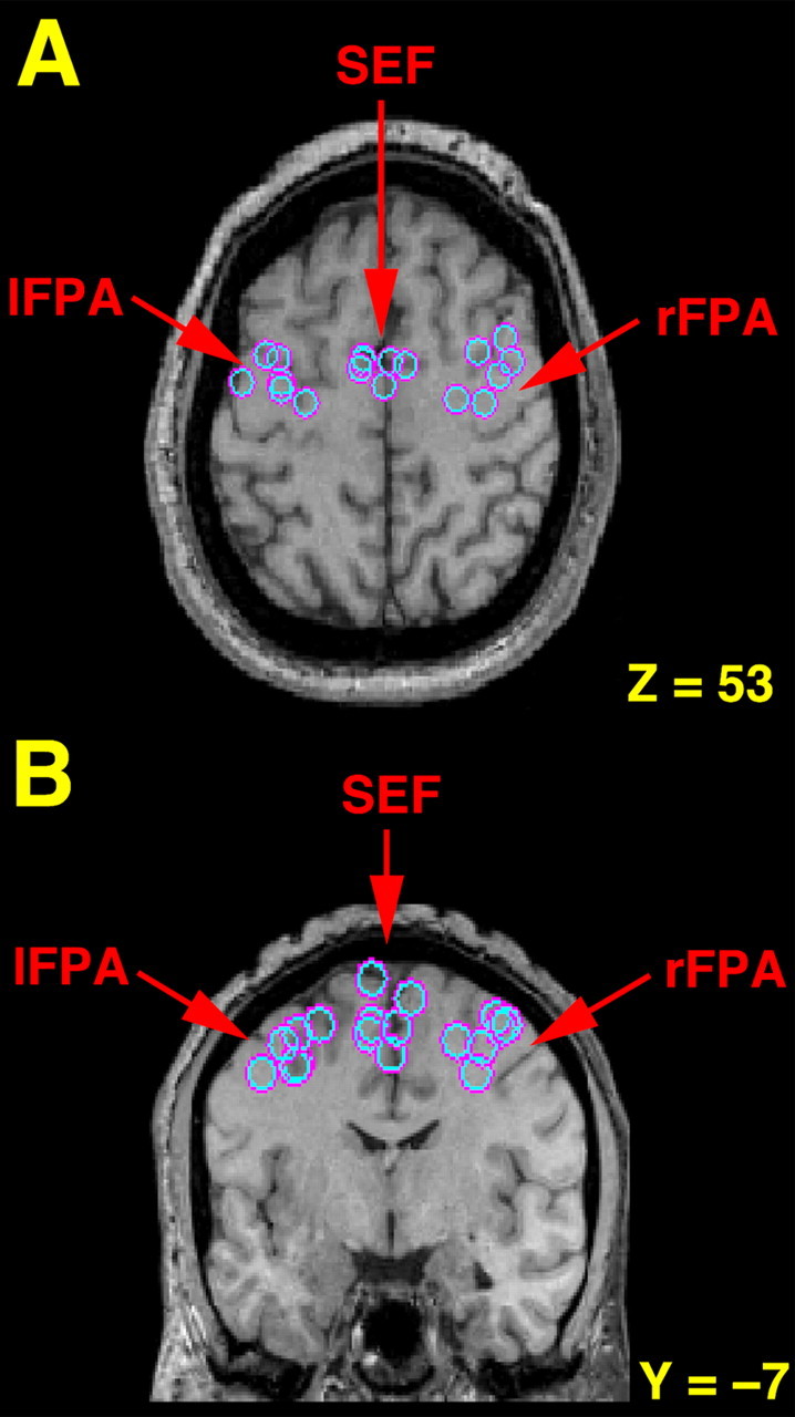
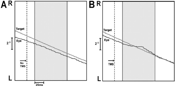
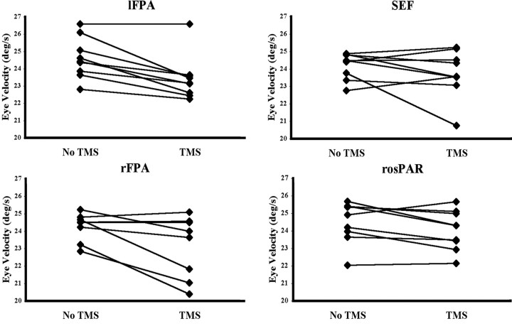
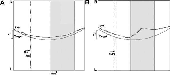
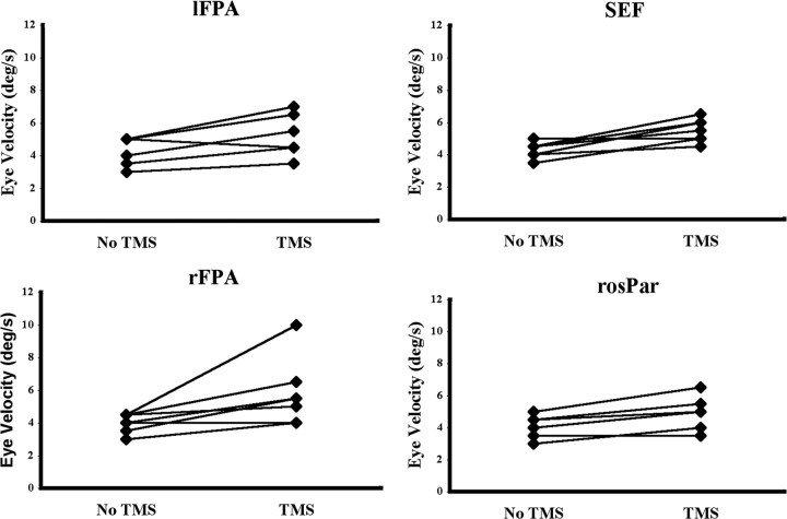
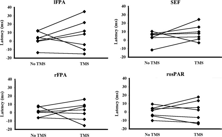
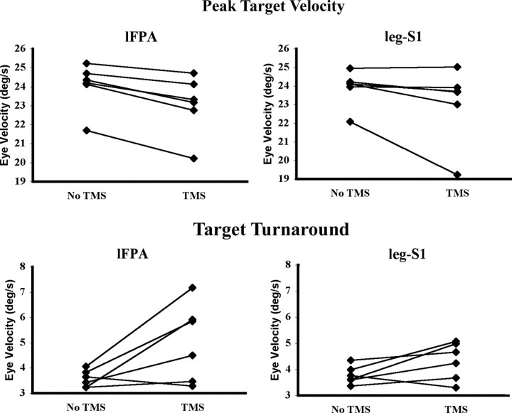
Both the frontal eye fields (FEFs) and supplementary eye fields (SEFs) are known to be involved in smooth pursuit eye movements. It has been shown recently that stimulation of the smooth-pursuit area of the FEF [frontal pursuit area (FPA)] in monkey increases the pursuit response to unexpected changes in target motion during pursuit. In the current study, we applied transcranial magnetic stimulation (TMS) to the FPA and SEF in humans during sinusoidal pursuit to assess its effects on the pursuit response to predictable, rather than unexpected, changes in target motion. For the FPA, we found that TMS applied immediately before the target reversed direction increased eye velocity in the new direction, whereas TMS applied in mid-cycle, immediately before the target began to slow, decreased eye velocity. For the SEF, TMS applied at target reversal increased eye velocity in the new direction but had no effect on eye velocity when applied at mid-cycle. TMS of the control region (leg region of the somatosensory cortex) did not affect eye velocity at either point. Previous stimulation studies of FPA during pursuit have suggested that this region is involved in controlling the gain of the transformation of visual signals into pursuit motor commands. The current results suggest that the gain of the transformation of predictive signals into motor commands is also controlled by the FPA. The effect of stimulation of the SEF is distinct from that of the FPA and suggests that its role in sinusoidal pursuit is primarily at the target direction reversal.
Figures
Similar articles
-
Enhancement of multiple components of pursuit eye movement by microstimulation in the arcuate frontal pursuit area in monkeys.J Neurophysiol. 2002 Feb;87(2):802-18. doi: 10.1152/jn.00409.2001. J Neurophysiol. 2002. PMID: 11826048 Free PMC article.
-
Role of arcuate frontal cortex of monkeys in smooth pursuit eye movements. I. Basic response properties to retinal image motion and position.J Neurophysiol. 2002 Jun;87(6):2684-99. doi: 10.1152/jn.2002.87.6.2684. J Neurophysiol. 2002. PMID: 12037171 Free PMC article.
-
The role of frontal pursuit area in interaction between smooth pursuit eye movements and attention: A TMS study.J Vis. 2021 Mar 1;21(3):11. doi: 10.1167/jov.21.3.11. J Vis. 2021. PMID: 33683288 Free PMC article.
-
The neuronal basis of on-line visual control in smooth pursuit eye movements.Vision Res. 2015 May;110(Pt B):257-64. doi: 10.1016/j.visres.2014.06.008. Epub 2014 Jul 1. Vision Res. 2015. PMID: 24995378 Free PMC article. Review.
-
The vestibular-related frontal cortex and its role in smooth-pursuit eye movements and vestibular-pursuit interactions.J Vestib Res. 2006;16(1-2):1-22. J Vestib Res. 2006. PMID: 16917164 Free PMC article. Review.
Cited by
-
Inactivation and stimulation of the frontal pursuit area change pursuit metrics without affecting pursuit target selection.J Neurophysiol. 2011 Jul;106(1):347-60. doi: 10.1152/jn.00669.2010. Epub 2011 Apr 27. J Neurophysiol. 2011. PMID: 21525365 Free PMC article.
-
Evaluation of the smooth pursuit tests in multiple sclerosis patients.J Neurol. 2011 Oct;258(10):1795-800. doi: 10.1007/s00415-011-6014-0. Epub 2011 Mar 29. J Neurol. 2011. PMID: 21445600
-
Anticipatory smooth pursuit eye movements scale with the probability of visual motion: The role of target speed and acceleration.J Vis. 2025 Jan 2;25(1):2. doi: 10.1167/jov.25.1.2. J Vis. 2025. PMID: 39752177 Free PMC article.
-
Neural activity in the frontal pursuit area does not underlie pursuit target selection.Vision Res. 2011 Apr 22;51(8):853-66. doi: 10.1016/j.visres.2010.10.010. Epub 2010 Oct 21. Vision Res. 2011. PMID: 20970442 Free PMC article.
-
Human middle temporal cortex, perceptual bias, and perceptual memory for ambiguous three-dimensional motion.J Neurosci. 2010 Jan 13;30(2):760-6. doi: 10.1523/JNEUROSCI.4171-09.2010. J Neurosci. 2010. PMID: 20071541 Free PMC article.
References
-
- Barnes GR, Donelan SF (1999) The remembered pursuit task: evidence for segregation of timing and velocity storage in predictive oculomotor control. Exp Brain Res 129: 57–67. - PubMed
-
- Bullmore ET, Brammer MJ, Williams SCR, Rabe-Hesketh S, Janot N, David AS, Mellers JDC, Howard R, Sham P (1996) Statistical methods of estimation and inference for functional MR image analysis. Magn Reson Med 35: 261–277. - PubMed
-
- Cracco RQ, Cracco JB, Maccabee PJ, Amassian VE (1999) Cerebral function revealed by transcranial magnetic stimulation. J Neurosci Methods 86: 209–219. - PubMed
-
- Culham JC, Kanwisher NG (2001) Neuroimaging of cognitive functions in human parietal cortex. Curr Opin Neurobiol 11: 157–163. - PubMed
Publication types
MeSH terms
LinkOut - more resources
Full Text Sources