Comparative Study
. 2006 Jan 11;26(2):550-8.
doi: 10.1523/JNEUROSCI.3859-05.2006.

Inhibitors of differentiation and DNA binding (Ids) regulate Math1 and hair cell formation during the development of the organ of Corti

Affiliations
Comparative Study

Inhibitors of differentiation and DNA binding (Ids) regulate Math1 and hair cell formation during the development of the organ of Corti

Jennifer M Jones et al. J Neurosci. .

Abstract

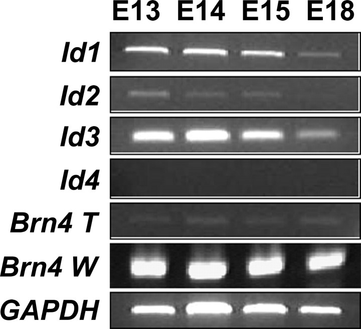
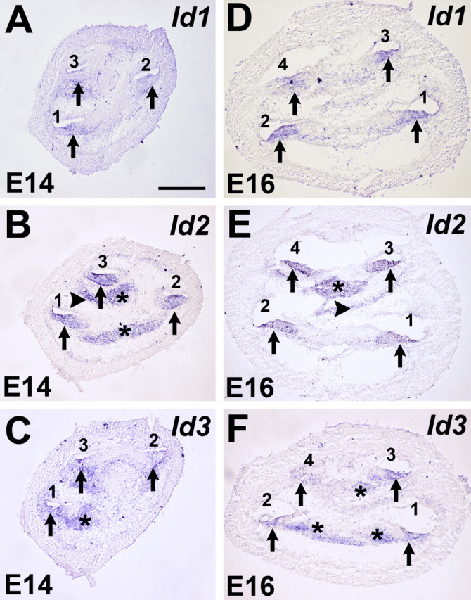
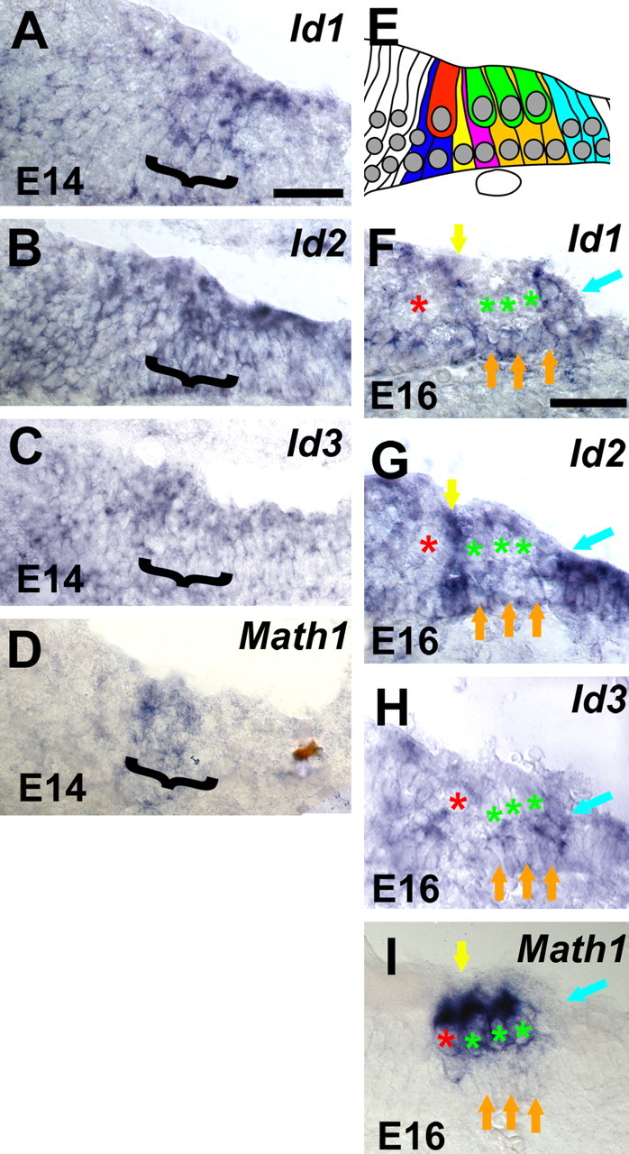
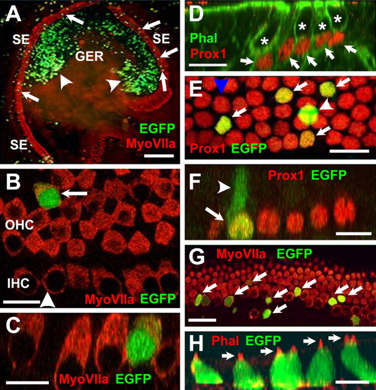
The basic helix-loop-helix (bHLH) transcription factor Math1 (also called Atoh1) is both necessary and sufficient for hair cell development in the mammalian cochlea (Bermingham et al., 1999; Zheng and Gao, 2000). Previous studies have demonstrated that a dynamic pattern of Math1 expression plays a key role in regulating the number and position of mechanosensory hair cells. However, the factors that regulate the temporal and spatial expression of Math1 within the cochlea are unknown. The bHLH-related inhibitors of differentiation and DNA binding (Id) proteins are known to negatively regulate many bHLH transcription factors, including Math1, in a number of different systems. Therefore, Id proteins are good candidates for regulating Math1 in the cochlea. Results from PCR and in situ hybridization indicate that Id1, Id2, and Id3 are expressed within the cochlear duct in a pattern that is consistent with a role in regulation of hair cell development. In particular, expression of Ids and Math1 overlapped in cochlear progenitor cells before cellular differentiation, but a specific downregulation of Id expression was observed in individual cells that differentiated as hair cells. In addition, progenitor cells in which the expression of Ids was maintained during the time period for hair cell differentiation were inhibited from developing as hair cells. These results indicate a key role for Ids in the regulation of expression of Math1 and hair cell differentiation in the developing cochlea.


Figures

Figure 1.
Figure 1.
Expression of Id mRNAs is downregulated as the organ of Corti matures. Total RNA was isolated from cells before hair cell differentiation (E13), at the onset of hair cell differentiation (E14 and E15), and near the end of embryonic development (E18), and expression of Ids was detected by PCR. mRNAs for Id1, Id2, and Id3 are expressed at E13, E14, and E15 but are downregulated by E18. In contrast, Id4 is not expressed at any time point. Amplification of GAPDH was used to control for equal loading of cDNA in each reaction. To ensure that the cochlear samples were not contaminated by surrounding mesenchymal tissue, Brn4, a molecule that is not expressed in the developing cochlear epithelium but is expressed in adjacent mesenchyme, was amplified from cochlear epithelial samples (Brn4 T) and from samples containing both cochlear epithelium and surrounding mesenchyme (Brn4 W). Although there is a strong band for Brn4 in the Brn4 W samples at each time point, only minimal Brn4 was amplified from the Brn4 T sample, indicating minimal contamination by mesenchymal cells in the isolated epithelial samples.
Figure 2.
Figure 2.
Expression of Id1, Id2, and Id3 in the developing cochlea. A–F, In situ hybridizations for Id1, Id2, and Id3 in mid-modiolar cochlear sections at E14 (A–C) and E16 (D–F). At E14, Id1 (A), Id2 (B), and Id3 (C) are broadly expressed in the floor of the cochlear duct (arrows) in each of the three cochlear turns, basal (1), middle (2), and apical (3). Expression of Id2 and Id3 was also observed in the developing spiral ganglion (B, C, asterisks), and Id2 expression was observed in some mesenchymal cells (B, arrowhead). At E16, expression of Id1 (D), Id2 (E), and Id3 (F) was still present in along the length of the cochlea duct, which has grown to four turns (numbered 1–4 as in A). Expression of Id2 and Id3 was also still present in the spiral ganglion (E and F, asterisks) and in some mesenchymal cells (E, arrowhead). Scale bar: (in A) B–F, 250 μm.
Figure 3.
Figure 3.
Id expression is downregulating in developing hair cells. High-magnification images of Id1, Id2, Id3, and Math 1 expression in the basal cochlear turn from mid-modiolar sections at E14 and E16. Color has been removed to provide enhanced contrast. A–C, At E14, Id1 (A), Id2 (B), and Id3 (C) are broadly expressed within the developing cochlea, including progenitor cells that will develop as the organ of Corti (bracketed region). D, At the same time point, Math 1 (bracket) is also expressed as a subset of the cells that express Ids. At this stage, Math 1 expression is an indicator of the prosensory region. E, Schematic illustration of individual cell types within the developing organ of Corti at E16. A single inner hair cell (red) and three outer hair cells (green) can be identified closer to the lumenal surface of the epithelium. Specific supporting cell types, including inner phalangeal cells (blue), inner pillar cells (yellow), outer pillar cells (magenta), Deiter's cells (orange), and Hensen's cells (blue) can also be identified based on nuclear position. At E16, expression of Id1 (F), Id2 (G), and Id3 (H) persists in supporting cell types, but there is a marked decrease in the expression of all three Ids in developing hair cells. In each panel, inner and outer hair cell nuclei are indicated with red and green asterisks. Maintained expression of Id1, Id2, and Id3 can be clearly observed in pillar cells (yellow arrows), Deiter's cells (orange arrows), and Hensen's cells (blue arrows). In contrast, by this stage, expression of Math 1 is restricted to inner (red asterisk) and outer (green asterisks) hair cells. All supporting cell types (arrows as in F) are negative for Math 1. Scale bars: (in A) B, C, 25 μm; (in E) F, G, 25 μm.
Figure 4.
Figure 4.
Transfection of progenitor cells in cochlear explant cultures. A, Low-magnification view of an explant culture electroporated at E13 with an EGFP expression plasmid and maintained in vitro for 6 d. Hair cells within the sensory epithelium (SE) have been labeled with anti-myosin VIIa (red), and transfected cells expressing EGFP are green. EGFP-positive cells (green) are present in the GER (arrowheads) and the SE (arrows). B, High-magnification view of the sensory epithelium in an EGFP-transfected explant. The single row in inner hair cells (IHC) and, in this case, four rows of outer hair cells (OHC) are positive for myosin VIIa (red). A single transfected cell (green) has developed as an outer hair cell (arrow). The arrowhead indicates the plane of Z-section in C. C, Confocal Z-stack cross section along the plane indicated by the arrowhead in B, illustrating hair cell morphology in the transfected cell (green). D, Prox1 is a marker for supporting cells in the organ of Corti. Confocal Z-stack of a P0 mouse cochlea. Prox1 expression is illustrated in red. Filamentous actin is labeled with phalloidin in green. Nuclei of supporting cells (arrows) are positive for Prox1 (red), whereas nuclei of hair cells (asterisks) are negative for Prox1. E, High-magnification view of the supporting cell nuclear layer in an explant transfected with EGFP. Four transfected cells (green, arrows) are also positive for Prox1. A single additional cell (arrowhead) is also transfected but appears to be located at a more lumenal position within the epithelium and is not Prox1 positive. This cell has probably developed as a hair cell. The blue arrowhead indicates plane of Z-section in F. F, Confocal Z-stack cross section through the plane indicated by the blue arrowhead in E. The single transfected Prox1-positive cell (arrow) has a morphology that is consistent with a supporting cell, including an apical projection (arrowhead) that extends to the lumenal surface. G, Overexpression of Math 1 induces development of hair cells in the sensory epithelium. Virtually all progenitor cells transfected with Math 1 (green, arrows) are positive for myosin VIIa (red), indicating that they have developed as hair cells. H, Math 1-transfected cells also develop stereociliary bundles. Confocal Z-stack cross section through a group of transfected progenitor cells (green). Stereociliary bundles on each hair cell (arrows) are visualized by phalloidin labeling of filamentous actin (red). Scale bars: A, 250 μm; B, 20 μm; C, 10 μm; D, 20 μm; E, 20 μm; F, 20 μm; G, 20 μm; H, 10 μm.
Figure 5.
Figure 5.
Overexpression of Id3 inhibits hair cell differentiation but does not prevent supporting cell development in the sensory epithelium. A, Sensory epithelium in an explant transfected with Id3 (green; indicated as Id3-EGFP). Inner and outer hair cells are labeled with myosin VIIa (red). Two transfected cells (arrows) are present. Each cell is negative for myosin VIIa and has a morphology that is consistent with a supporting cell. B, Confocal Z-stack cross section of the two Id3-transfected cells in A. Each has a cell body (asterisks) located near the basement membrane and a lumenal projection (arrowheads). C, Confocal Z-stack view of a cell transfected with Id3 (green). Hair cells in the same image have been labeled with myosin VIIa in red. Note the basal position of the cell body (asterisk) and the lumenal projection (arrowhead). D, Surface view of the sensory epithelium in an explant labeled with the supporting cell marker Jagged1 (Jag1; red). Hair cells appear as black circles (asterisks), whereas interdigitating supporting cells are red. E, The same image as that in D, with three Id3-transfected cells illustrated in green (arrows). Note that each transfected cell is also positive for Jag1. F, Confocal Z-stack cross section of a cell transfected with Id3 (green). Note the large cell body located adjacent to the basement membrane and the thin extension projecting toward the lumenal surface. Also, a hair cell (asterisk) is located adjacent to the apical projection of this supporting cell, suggesting that this is a Deiter cell. G, Surface view of the sensory epithelium in an explant labeled with the supporting cell marker Prox1 (red). A single Id3-transfected cell is illustrated (green). The nucleus of this cell is positive for Prox1, making it appear yellow (arrow). The cell also extends a lumenal projection. The arrowhead indicates the plane of section for the Z-stack in H. H, Confocal Z-stack cross section of the cell in G, illustrating the lumenal projection. Note the yellow nucleus (arrow) indicating expression of Prox1. Based on the shape and position of this cell, it is most likely a third-row Deiter cell. I, A single Id3-transfected cell (green) that is also positive for p75NTR (red). This cell was located in the row of inner pillar cells, which all express p75NTR at their lumenal surfaces, giving rise to a stripe of p75NTR expression (arrow). J, Confocal Z-stack cross section of the same cell as in I. The morphology of this cell is consistent with a pillar cell. Scale bars: A, 20 μm; B–E, 10 μm; F, 5 μm; G, 10 μm; H–J, 5 μm.

Similar articles

Cited by

References

    1. Andres-Barquin PJ, Hernandez MC, Israel MA (2000) Id genes in nervous system development. Histol Histopathol 15: 603–618. - PubMed
    1. Anniko M (1983) Cytodifferentiation of cochlear hair cells. Am J Otolaryngol 4: 375–388. - PubMed
    1. Benezra R, Davis RL, Lockshon D, Turner DL, Weintraub H (1990) The protein Id: a negative regulator of helix-loop-helix DNA binding proteins. Cell 61: 49–59. - PubMed
    1. Bermingham NA, Hassan BA, Price SD, Vollrath MA, Ben-Arie N, Eatock RA, Bellen HJ, Lysakowski A, Zoghbi HY (1999) Math1: an essential gene for the generation of inner ear hair cells. Science 284: 1837–1841. - PubMed
    1. Bermingham-McDonogh O, Stone J, Hume C, Oesterle E (2005) Developmental expression of the homeobox transcription factor Prox1 in the murine organ of Corti. Assoc Res Otolaryngol Abstr 1353.

Publication types

MeSH terms

Substances