. 2006 Jan 11;26(2):597-608.
doi: 10.1523/JNEUROSCI.4493-05.2006.

GABAergic signaling at mossy fiber synapses in neonatal rat hippocampus

Affiliations

GABAergic signaling at mossy fiber synapses in neonatal rat hippocampus

Victoria F Safiulina et al. J Neurosci. .

Abstract

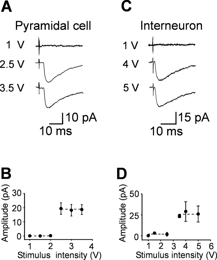
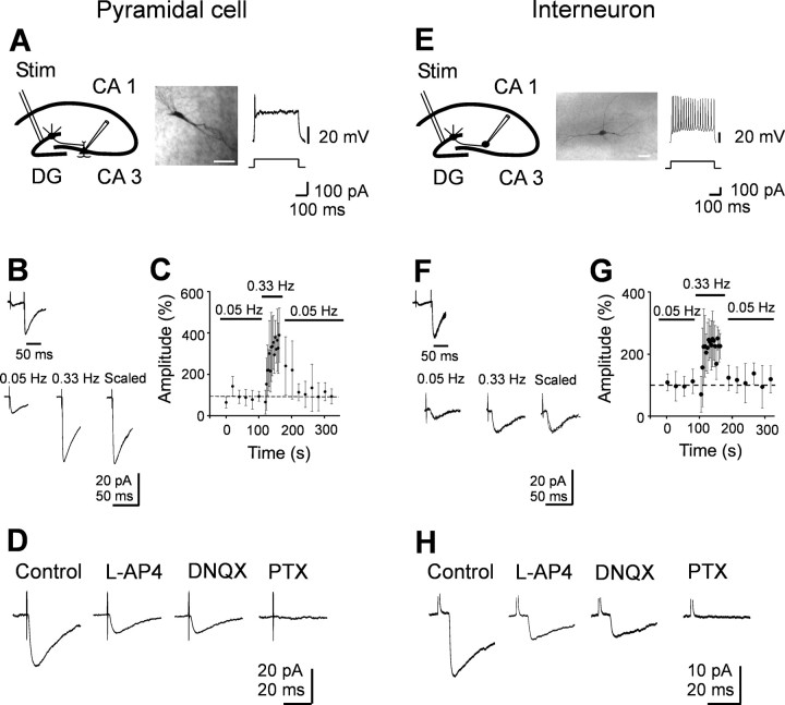
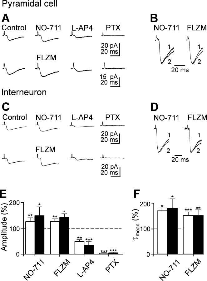
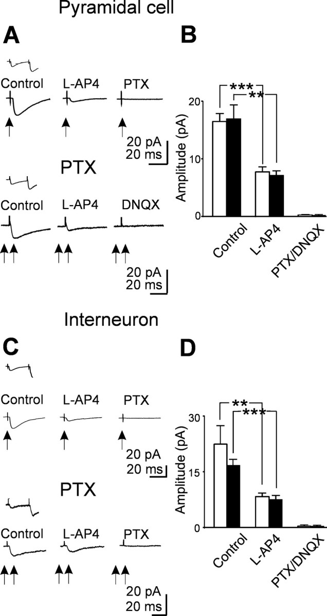
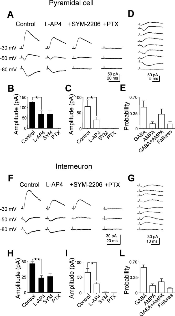
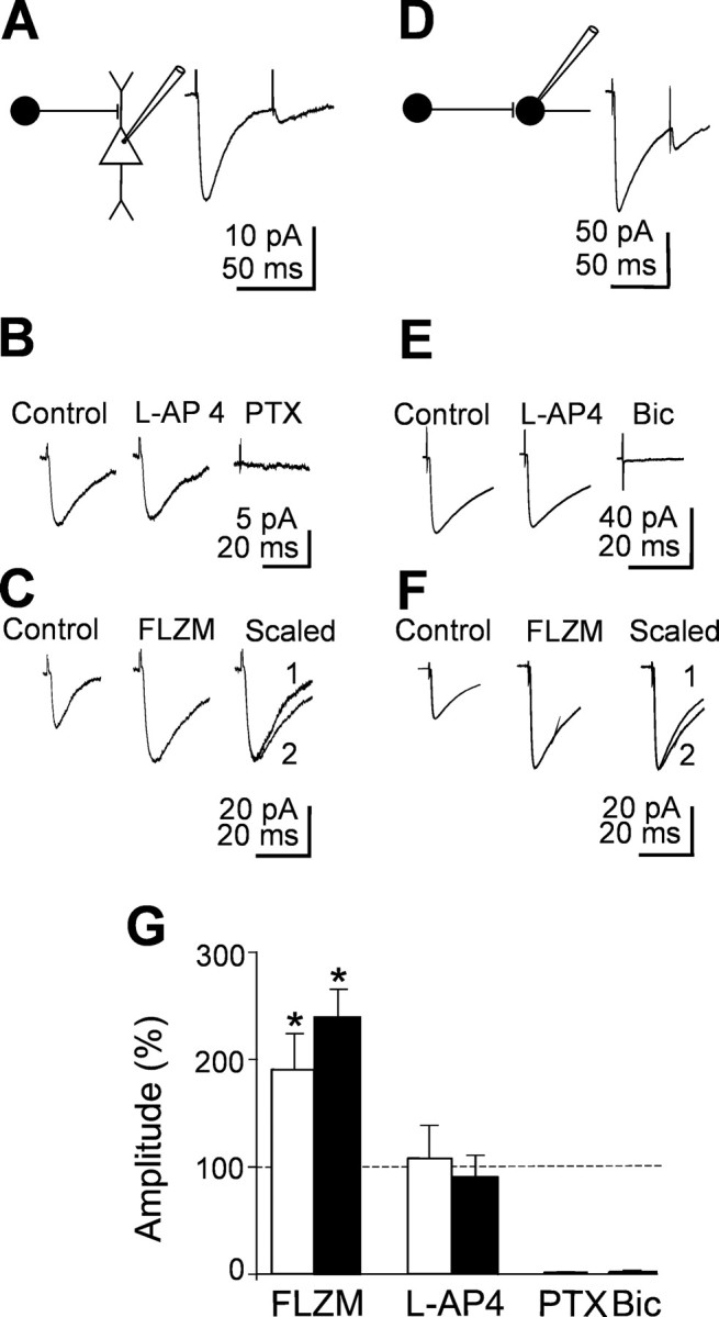
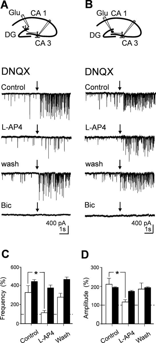
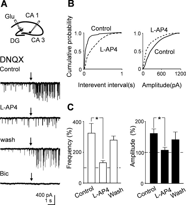
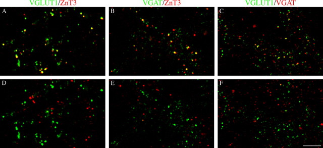
In the adult rat hippocampus, granule cell mossy fibers (MFs) form excitatory glutamatergic synapses with CA3 principal cells and local inhibitory interneurons. However, evidence has been provided that, in young animals and after seizures, the same fibers can release in addition to glutamate GABA. Here we show that, during the first postnatal week, stimulation of granule cells in the dentate gyrus gave rise to monosynaptic GABAA-mediated responses in principal cells and in interneurons. These synapses were indeed made by MFs because they exhibited strong paired-pulse facilitation, high sensitivity to the metabotropic glutamate receptor agonist l-AP-4, and short-term frequency-dependent facilitation. MF responses were potentiated by blocking the plasma membrane GABA transporter GAT-1 with NO-711 or by allosterically modulating GABAA receptors with flurazepam. Chemical stimulation of granule cell dendrites with glutamate induced barrages of GABAA-mediated postsynaptic currents into target neurons. Furthermore, immunocytochemical experiments demonstrated colocalization of vesicular GABA transporter with vesicular glutamate transporter-1 and zinc transporter 3, suggesting that GABA can be taken up and stored in synaptic vesicles of MF terminals. Additional fibers releasing both glutamate and GABA into principal cells and interneurons were recruited by increasing the strength of stimulation. Both the GABAergic and the glutamatergic component of synaptic currents occurred with the same latency and were reversibly abolished by l-AP-4, indicating that they originated from the MFs. GABAergic signaling may play a crucial role in tuning hippocampal network during postnatal development. Low-threshold GABA-releasing fibers may undergo elimination, and this may occur when GABA shifts from the depolarizing to the hyperpolarizing direction.


Figures

Figure 1.
Figure 1.
Unitary synaptic currents evoked in principal cells and interneurons by minimal stimulation of granule cells in the dentate gyrus. Different stimulus intensities were used to evoke synaptic currents in a CA3 pyramidal cell (A) at P3 or in an interneuron (C) at P4. Each trace is the average of 15-20 responses. Holding potential was -70 mV. The peak amplitude of synaptic currents represented in A and C is plotted against different stimulus intensities in B and D, respectively. Note the all-or-none appearance of synaptic currents with increasing stimulus intensities. Error bars indicate SEM. The dashed lines connect the mean values of individual points within the same groups.
Figure 2.
Figure 2.
Minimal stimulation of granule cells in the dentate gyrus evokes GABAA-mediated monosynaptic responses in CA3 pyramidal cells and interneurons. A, E, Left, Diagrams of the hippocampus showing a CA3 pyramidal cell (A) and an interneuron (E) with converging inputs from MFs. Stim, Stimulating electrode. Middle, Neurons labeled with biocytin. Scalebar, 50 μm. Right, Representative traces showing the typical firing pattern of a CA3 pyramidal cell and an interneuron at P3 in response to long depolarizing current pulses. B, F, Paired stimuli delivered at 50 ms interval to granule cells in the dentate gyrus evoked at -70 mV synaptic responses exhibiting strong paired-pulse facilitation (average of 50 individual responses). Bottom, Mean amplitude of synaptic responses (average of 15 individual traces) evoked by stimulation of granule cells in the dentate gyrus at 0.05 and 0.33 Hz. On the right, the two responses are normalized and superimposed. C, G, The mean amplitude of synaptic currents evoked in three pyramidal cells (C) and in three interneurons (G) at 0.05 and 0.33 Hz (bars) is plotted against time. Note the slow buildup of facilitation of synaptic responses at 0.33 Hz that completely reversed to control values after returning to 0.05 Hz stimulation. Error bars represent SEM. D, H, Synaptic responses from another pyramidal cell and interneuron before and during application of l-AP-4 (10 μm) and l-AP-4 plus DNQX (20 μm). Addition of picrotoxin (PTX; 100 μm) completely abolished synaptic currents. Each response is the average of 20 individual traces (including failures). Note that GABAA-mediated synaptic currents were depressed by l-AP-4 but were unaffected by DNQX.
Figure 3.
Figure 3.
GABAA-mediated MF responses are modulated by the GABA transporter GAT-1 and by flurazepam. A, Average of 20 individual responses (including failures) recorded from a CA3 pyramidal cell before and during application of the GAT-1 antagonist NO-711 (10 μm; top traces) or flurazepam (FLZM; 3 μm; bottom traces). The currents were reduced in amplitude by l-AP-4 (10 μm) and were blocked by picrotoxin (PTX; 100 μm). B, Normalized responses obtained in the absence (1) or in the presence (2) of NO-711 and FLZM, respectively. Note the slowdown of the deactivation kinetics of synaptic currents with these drugs. C and D refer to average traces obtained from an interneuron (legend as in A and B). The amplitude (E) and τmean (F) of synaptic currents obtained from five pyramidal cells (white columns) and six interneurons (black columns) recorded in different experimental conditions as indicated and normalized to the respective controls (dashed lines are shown). In this and the following figures, *p < 0.05, **p < 0.01, and ***p < 0.001.
Figure 4.
Figure 4.
Mossy fibers releasing glutamate into target neurons can be recruited by increasing the strength of stimulation. A, Top traces, Average of 20 individual traces (successes plus failures) evoked in a principal cell (at 33°C and at -70mV; arrow) by minimal stimulation of the granule cell in the dentate gyrus in control and in the presence of l-AP-4 and l-AP-4 plus picrotoxin (PTX). The inset above the traces shows average pair responses obtained in control. Bottom traces, Average of 20 responses evoked in another neuron in the presence of picrotoxin (100 μm) by increasing stimulus strength (from 3 to 13 V; double arrows). B, Summary data from eight principal cells showing the mean amplitude of the GABAergic (white columns) and glutamatergic (black columns) components of synaptic currents evoked in different experimental conditions. C, Average traces recorded from an interneuron (same legend as in A). D, Summary data from eight interneurons showing the mean amplitude of the GABAergic (white columns) and glutamatergic (black columns) components of synaptic currents obtained in the same experimental conditions as in B.
Figure 5.
Figure 5.
Both glutamatergic and GABAergic currents can be evoked in principal cells and interneurons by stimulating the granule cells in the dentate gyrus at room temperature. A, Average responses evoked at three different holding potentials in a CA3 pyramidal cell recorded with a low chloride intrapipette solution (ECl of -90 mV). Note the biphasic currents at -50 mV and the isolated GABAergic and glutamatergic components at -30 and -80 mV, respectively. The two components were reduced in amplitude by l-AP-4 and were selectively blocked by the AMPA and GABAA receptor blockers SYM 2206 (20 μm) and picrotoxin (PTX; 100 μm), respectively. B, C, Summary data (n = 5) showing the mean amplitude of GABAergic (black columns) and glutamatergic (white columns) currents in control and in the presence of l-AP-4, l-AP-4 plus SYM 2206, and l-AP-4 plus SYM 2206 and picrotoxin. D, Individual traces from one single cell recorded at -50 mV showing inward, outward, biphasic responses, and response failures. E, Each column represents the relative frequency of each type of response for three cells. F, G, Average traces recorded from an interneuron. H, I, L, Summary data from five interneurons (legend as in A-E).
Figure 6.
Figure 6.
l-AP-4-insensitive synaptic currents elicited in principal cells and interneurons by stimulation of the dentate gyrus. A, D, Left, Diagrams showing GABAergic interneurons impinging into a pyramidal cell (A) and into an interneuron (D). Right, Average of 20 individual responses (including failures) to paired stimuli. B, E, Average of 20 individual traces evoked in a pyramidal cell (B) or in an interneuron (E) in control and in the presence of l-AP-4 and l-AP-4 plus picrotoxin (PTX). Note the lack of effect of l-AP-4. C, F, Average of 20 individual traces from another pyramidal cell (C) and interneuron (F) obtained in control or in the presence of flurazepam (FLZM). On the right, the two normalized traces are superimposed. G, Summary data showing the amplitude of synaptic currents obtained from four pyramidal cells (white columns) and four interneurons (black columns) in different experimental conditions (as indicated) and normalized to the respective controls (dashed lines). Bic, Bicuculline.
Figure 7.
Figure 7.
Pressure application of glutamate to stratum moleculare evokes l-AP-4-sensitive GABAergic currents in principal cells. A, Diagram showing the location of the patch and puff pipette. Glutamate was applied by pressure from a pipette positioned into stratum moleculare, in the presence of DNQX (20 μm). Individual traces obtained in control, in the presence of l-AP-4, after washing l-AP-4, and in the presence of bicuculline (Bic; 20 μm) before and after (arrows) application of glutamate. B, Same as A, but the puff pipette was positioned close to the hilus. Note that, in this case, glutamate-evoked GABAergic currents were poorly affected by l-AP-4. Mean frequency (C) and amplitude (D) values of GABAergic currents (normalized to pre-glutamate values) evoked by pressure application of glutamate to stratum moleculare (white; n = 6) or to the hilus (black; n = 3).
Figure 8.
Figure 8.
Pressure application of glutamate to stratum moleculare evokes l-AP-4-sensitive GABAergic currents in GABAergic interneurons. A, Diagram showing the location of the patch and puff pipette. Glutamate was applied by pressure from a pipette positioned into stratum moleculare, in the presence of DNQX (20 μm). Individual traces obtained in control, in the presence of l-AP-4, after washing l-AP-4, and in the presence of bicuculline (20 μm) before and after (arrows) application of glutamate. B, Cumulative interevent and amplitude distribution histograms of glutamate-evoked synaptic currents recorded from the cell shown in A in control (continuous lines) and during application of l-AP-4 (dashed lines). C, Mean frequency (white columns) and amplitude (black columns) values of GABAA-mediated currents (normalized to pre-glutamate values) evoked in five GABAergic interneurons by pressure application of glutamate to stratum moleculare.
Figure 9.
Figure 9.
Immunocytochemical evidence for the expression of VGAT in MF terminals. VGAT expression in CA3 VGLUT1- and ZnT3-positive fibers at P5. A, VGLUT1/ZnT3-positive puncta. B, C, VGLUT1- and ZnT3-positive puncta coexpress VGAT immunoreactivity in PL. D-F, Rotation of the red channel by 90° strongly reduces colocalization. SL, Stratum lucidum. Scale bar: (in F) A-F, 5 μm.

Similar articles

Cited by

References

    1. Acsády L, Kamondi A, Sik A, Freund T, Buzsaki G (1998) GABAergic cells are the major postsynaptic targets of mossy fibers in the rat hippocampus. J Neurosci 18: 3386-3403. - PMC - PubMed
    1. Allen C, Stevens CF (1994) An evaluation of causes for unreliability of synaptic transmission. Proc Natl Acad Sci USA 91: 10380-10383. - PMC - PubMed
    1. Amaral DG, Dent JA (1981) Development of the mossy fibers of the dentate gyrus. I. A light and electron microscopic study of the mossy fibers and their expansions. J Comp Neurol 195: 51-86. - PubMed
    1. Bartos M, Vida I, Frotscher M, Geiger JR, Jonas P (2001) Rapid signaling at inhibitory synapses in a dentate gyrus interneuron network. J Neurosci 21: 2687-2698. - PMC - PubMed
    1. Bekkers JM (2005) Presynaptically silent GABA synapses in hippocampus. J Neurosci 25: 4031-4039. - PMC - PubMed

Publication types

MeSH terms

Substances

LinkOut - more resources