Comparative Study
. 2006 Jan 11;26(2):644-54.
doi: 10.1523/JNEUROSCI.3861-05.2006.

Impaired glutamate transport in a mouse model of tau pathology in astrocytes

Affiliations
Comparative Study

Impaired glutamate transport in a mouse model of tau pathology in astrocytes

Deepa V Dabir et al. J Neurosci. .

Abstract

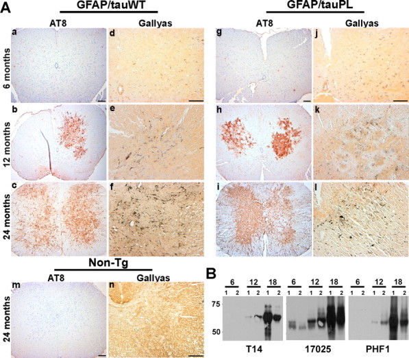
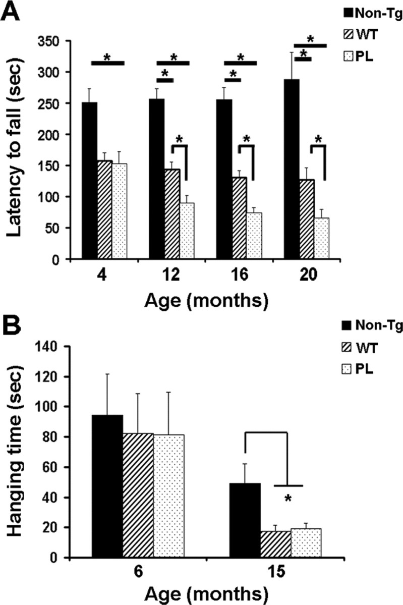
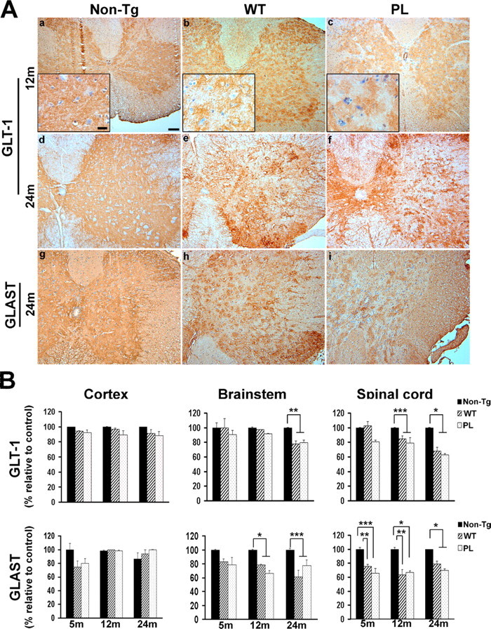
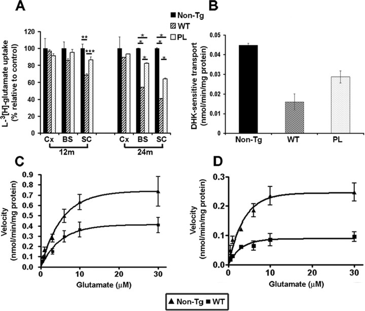
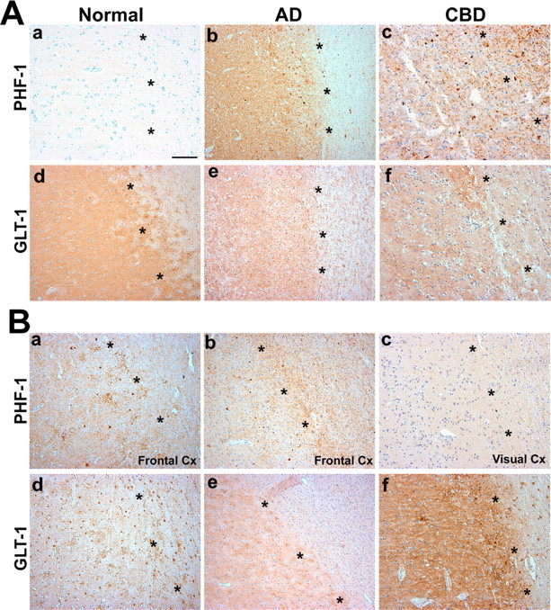
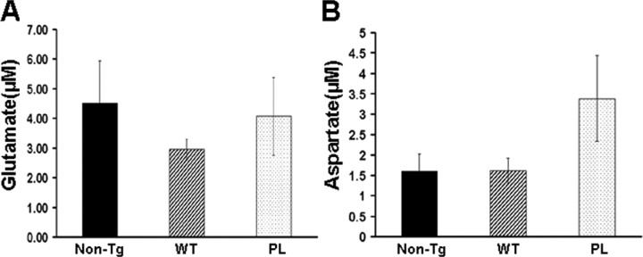
Filamentous tau inclusions in neurons and glia are neuropathological hallmarks of tauopathies. The discovery of microtubule-associated protein tau gene mutations that are pathogenic for a heterogenous group of neurodegenerative disorders, called frontotemporal dementia and parkinsonism linked to chromosome-17 (FTDP-17), directly implicate tau abnormalities in the onset/progression of disease. Although the role of tau pathology in neurons in disease pathogenesis is well accepted, the contribution of glial pathology is essentially unknown. We recently generated a transgenic (Tg) mouse model of tau pathology in astrocytes by expressing the human tau protein under the control of the glial fibrillary acidic protein (GFAP) promoter. Both wild-type and FTDP-17 mutant GFAP/tau Tg animals manifest an age-dependent accumulation of tau inclusions in astrocytes that resembles the pathology observed in human tauopathies. We further demonstrate that both strains of Tg mice manifest compromised motor function that correlates with altered expression of the glial glutamate-aspartate transporter and occurs before the development of tau pathology. Subsequently, the Tg mice manifest additional deficits in neuromuscular strength that correlates with reduced expression of glutamate transporter-1 (GLT-1) and occurs concurrent with tau inclusion pathology. Reduced GLT-1 expression was associated with a progressive decrease in sodium-dependent glutamate transport capacity. Reductions in GLT-1 expression were also observed in corticobasal degeneration, a tauopathy with prominent pathology in astrocytes. Less robust changes were observed in Alzheimer's disease in which neuronal tau pathology predominates. Thus, these Tg mice recapitulate features of astrocytic pathology observed in tauopathies and implicate a role for altered astrocyte function in the pathogenesis of these disorders.


Figures

Figure 1.
Figure 1.
Astrocyte-specific regional human tau expression in GFAP/tauWT and GFAP/tauP301L Tg mice. A, Western blot analysis of soluble tau protein extracted from cortex, brainstem, and spinal cord samples of 2-month-old control (non-Tg), GFAP/tauP301L (PL), and GFAP/tauWT Tg (WT) mice. Ten micrograms of soluble protein extract was resolved by SDS-PAGE and immunoblotted with T14, a monoclonal antibody (mAb) specific for human tau. Both GFAP/tauWT and GFAP/tauP301L Tg lines show highest levels of human tau protein expression in the spinal cord, whereas the lowest levels are detected in the cortex. The GFAP/tauWT Tg mouse expresses approximately fourfold higher levels of human tau protein than GFAP/tauP301L. Molecular weight standards are indicated to the left of each panel. B, Immunohistochemistry was performed with the human tau-specific mAb OT12 (B) on cortex, brainstem, and spinal cords of 2-month-old GFAP/tauWT (WT) and GFAP/tauP301L (PL) Tg mice. Robust tau staining is observed in astrocytes within the gray matter of tau Tg mice. Human tau was not detected in non-Tg mice (data not shown). Insets show tau staining in cells with astrocytic morphology. Scalebar: (in a) a, b, d, e, 200 μm; (inc) c, f, 500 μm; insets, 50 μm. C, Immunohistochemistry for GFAP was performed on the spinal cord of 6 month GFAP/tauWT, GFAP/tauP301L Tg, and non-Tg mice. There is prominent GFAP staining in spinal cord gray matter of GFAP/tauWT and GFAP/tauP301L Tg mice, but only limited GFAP staining is detected in spinal cord gray matter in the non-Tg animals. Scale bar, 100 μm.
Figure 2.
Figure 2.
Age-dependent accumulation of pathological tau inclusions in GFAP/tau Tg mice. A, Spinal cord from 6-, 12-, and 24-month-old GFAP/tauWT and GFAP/tauP301L mice were analyzed by immunohistochemistry with AT8, an antibody specific for tau phosphorylated at Ser202 and Thr205 (a-c, g-i, m) and Gallyas silver impregnation stain (d-f, j-l, n). There is an age-dependant accumulation of phosphorylated tau first detected at ∼12 months of age in both lines of tau Tg mice. Only a subset of the AT8-positive pathology is detected with Gallyas silver staining at this age. However, by 24 months, there is robust AT8 immunoreactivity and Gallyas-positive tau pathology in both astrocytic processes and cell soma of GFAP/tauWT and GFAP/tauP301L mice, which is not detected in 24-month-old non-Tg animals (m, n). The distribution and density of tau pathology is similar in the GFAP/tauWT and GFAP/tauP301L Tg mice. Scale bars: (in a) a-c, g-i, m, 100 μm; (in d) d-f, j-l, n, 100 μm. B, Immunoblot analysis of insoluble tau corresponding to 25 mg of starting wet tissue weight extracted from spinal cords of GFAP/tauP301L Tg mice at ages indicated and detected with T14, a human tau-specific mAb, 17025, a polyclonal antibody that detects both human and murine tau, and PHF-1, a mAb specific for tau phosphorylated at Ser396 and Ser404. At each age, two animals were analyzed. Insoluble tau protein is first detected in spinal cord and brainstem (data not shown) at 12 months of age. By 18 months of age, there is extensive insoluble and heavily aggregated tau pathology throughout the brainstem and spinal cord. Some variations in insoluble tau protein was observed between the two animals used at each age.
Figure 3.
Figure 3.
Motor impairment in GFAP/tau Tg mice. A, Both strains of tau Tg and non-Tg mice at the ages indicated were tested on the accelerating rotarod after 2 weeks of training. Latency to fall was recorded for each of 12 trials over 2 weeks. n = 12-14 animals per group at 4 months of age; n = 6-9 animals per group at 12, 16, and 20 months. Impaired performance was observed in both GFAP/tauWT (WT) and GFAP/tauP301L (PL) Tg mice relative to age-matched controls (one-way ANOVA, Bonferroni's post hoc test; *p < 0.001). At 12 months of age, the GFAP/tauP301L Tg mice also manifest impaired performance relative to the GFAP/tauWT Tg animals (one-way ANOVA, Bonferroni's post hoc test; *p < 0.001). Latency to fall, Mean of 12 trials. Error bars indicate SEM. B, GFAP/tau Tg and control mice at the ages indicated were assessed for neuromuscular strength using the wire-hang test. Animals were suspended from a wire, and the latency to fall from the wire was recorded as hanging time in seconds. Each mouse was tested four times over 2 d with an intertrial interval of 2 h. n = 11-14 mice per group at 6 months of age and 6-9 animals per group at 15 months. At 4 months of age, there was no difference in performance between both the strains of tau Tg and non-Tg mice. In contrast, both GFAP/tauWT (WT) and GFAP/tauP301L (PL) mice were significantly impaired at 15 months of age with 65% impairment relative to non-Tg mice (one-way ANOVA, Bonferroni's post hoc test; *p < 0.001). Hanging time, Mean of four trials. Error bars indicate SEM.
Figure 4.
Figure 4.
Decreased glial glutamate transporter expression in wild-type and mutant GFAP/tau Tg mice. A, Immunohistochemical analysis was performed on spinal cord sections from 12-month-old (a-c) and 24-month-old (d-i) GFAP/tau Tg and non-Tg mice, as indicated with antibodies to GLT-1 and GLAST. There is altered GLT-1 (a-c) and GLAST (data not shown) immunoreactivity relative to non-Tg animals at 12 months of age. Insets show higher magnification of patchy staining for GLT-1 in both strains of tau Tg mice compared with the diffuse gray matter staining observed in age-matched, non-Tg controls. At 24 months of age, there is progressive loss of both GLT-1 and GLAST immunoreactivity in both GFAP/tauWT and GFAP/tauP301L (PL) Tg animals. Scale bars: a-i, 100 μm; insets, 20 μm. B, Cortex, brainstem, and spinal cord from non-Tg, GFAP/tauWT, and GFAP/tauP301L Tg mice at 5, 12, and 24 months of age were extracted as described in Materials and Methods and immunoblotted with antibodies to GLT-1, GLAST, and actin (supplemental Fig. 2, available at www.jneurosci.org as supplemental material). Immunoblots were quantified with Multigauge version 2.3 software and normalized to actin levels. Data are presented as percentage of non-Tg age-matched control animals. At each time point, four animals per group were analyzed. GLAST expression was significantly decreased in both the brainstem (12 and 24 months) and spinal cord (5, 12, and 24 months) of both the strains of tau Tg mice relative to control animals (one-way ANOVA, Bonferroni's post hoc test; *p < 0.001; **p < 0.01; ***p < 0.05). In contrast, GLT-1 expression was only mildly decreased at 12 months of age in the GFAP/tauWT and GFAP/tauP301L Tg mice. However, at 24 months of age when the GFAP/tauWT and GFAP/tauP301L Tg mice manifest robust tau pathology, there was significant loss of GLT-1 protein expression in both the brainstem and spinal cord (one-way ANOVA, Bonferroni's post hoc test; *p < 0.001; **p < 0.01; ***p < 0.05). Representative immunoblots for human tau (T14), GLT-1, GLAST, EAAC1, and actin from brain regions indicated of 24-month-old animals are shown in supplemental Figure S2 (available at www.jneurosci.org as supplemental material).
Figure 5.
Figure 5.
Reduced sodium-dependent glutamate uptake in GFAP/tau Tg mice.A, l-[3H]Glutamate uptake was measured in P2 synaptosomal fractions prepared from cortex (Cx), brainstem (BS), and spinal cord (SC) of 12- and 24-month-old GFAP/tauWT (WT), GFAP/tauP301L (PL), and non-Tg mice. Initial rates of sodium-dependent uptake were expressed as nanomoles per minute per milligram of P2 synaptosomal protein extract. Data are presented as the percentage of glutamate uptake relative to age-matched non-Tg mice. At 12 months of age, there was a significant (32%) reduction in glutamate uptake capacity in the spinal cords of GFAP/tauWT Tg mice relative to the control animals (one-way ANOVA, Bonferroni's post hoc test; *p < 0.001). GFAP/tauP301L showed a smaller reduction (14%), but this was not statistically significant. At 24 months of age, there was an additional reduction in transport capacity in brainstem (WT, 46%; PL, 28%) and spinal cords (WT, 60%; PL, 36%) relative to the control. There were also significant differences in glutamate uptake in synaptosomal preparations from brainstem and spinal cord of 24-month-old GFAP/tauWT and GFAP/tauP301L Tg animals. Data represent mean from four mice analyzed in two independent experiments. Error bars indicate standard SEM (one-way ANOVA, Bonferroni's post hoc test; *p < 0.001; **p < 0.01; ***p < 0.05). B, l-[3H]Glutamate uptake into P2 synaptosomal fractions prepared from spinal cords of 24-month-old GFAP/tauWT, GFAP/tauP301L Tg, and age-matched control animals was measured in the presence and absence of 300 μm DHK, a selective GLT-1 inhibitor. The net sodium-dependent glutamate uptake values are provided that represent the difference in glutamate uptake in the presence and absence of DHK. Error bars represent SEM. C, D, Saturation isotherms for the high-affinity sodium-dependent transport of glutamate in spinal cord P2 synaptosomal fractions of GFAP/tauWT (WT) Tg and non-Tg controls at 12 (C) and 24 (D) months of age. Data represent the mean of four mice performed in two independent experiments. Error bars represent SEM. At 12 months of age, Vmax is reduced 43% relative to the non-Tg controls. By 24 months of age, Vmax is further reduced to 60% (one-way ANOVA, Bonferroni's post hoc test; p < 0.001). In contrast, there was no statistically significant difference in Km between the GFAP/tauWT Tg and non-Tg mice.
Figure 6.
Figure 6.
Loss of GLT-1 immunoreactivity in affected cortex of CBD patients. A, Adjacent sections from frontal cortex from patients with CBD (c, f), AD (b, e), and age-matched controls (a, d) were immunostained with PHF-1 (a-c) and GLT-1 (d-f). In cortex from control patients, there is little/no tau pathology (a), and GLT-1 shows diffuse cortical staining (d). In contrast, affected cortex from CBD patients shows robust tau pathology in both gray and white matter (c), with a marked loss of GLT-1 immunoreactivity in cortex (f). Occasional GLT-1-positive astrocytes are observed in the white matter. In frontal cortex from patients with AD, tau pathology is primarily restricted to the neocortex (b), with only mild and variable loss of GLT-1 immunoreactivity (e). B, Adjacent sections of frontal (a, b, d, e) and visual (c, f) cortex (Cx) from additional patients with CBD were immunostained with PHF-1 (a-c) and GLT-1 (d-f). Similar to A, there was robust tau pathology in both gray and white matter of affected cortex from CBD patients (A, B), with a marked loss of GLT-1 immunoreactivity in cortex (d, e). In contrast, in unaffected visual cortex, there is limited tau pathology (c), and, similar to control patients, GLT-1 shows diffuse cortical staining (f). The series of * symbols in each panel demarcate gray (to the left in each photomicrograph) from white matter. Scale bar, 500 μm
Figure 7.
Figure 7.
CSF glutamate (A) and aspartate (B) levels in GFAP/tau Tg mice. CSF samples from cisterna magna of 24-month-old GFAP/tauWT, GFAP/tauP301L Tg, and non-Tg mice were analyzed by HPLC for glutamate and aspartate. No significant differences in concentrations of CSF glutamate and aspartate were detected between both strains of tau Tg and control mice. The amino acids methionine, tryptophan, GABA, and asparagine were also analyzed, but no difference between groups was observed (data not shown). Six animals per group were used for the CSF analyses. Error bars indicate SEM.

Similar articles

Cited by

References

    1. Araque A, Carmignoto G, Haydon PG (2001) Dynamic signaling between astrocytes and neurons. Annu Rev Physiol 63: 795-813. - PubMed
    1. Arriza JL, Fairman WA, Wadiche JI, Murdoch GH, Kavanaugh MP, Amara SG (1994) Functional comparisons of three glutamate transporter subtypes cloned from human motor cortex. J Neurosci 14: 5559-5569. - PMC - PubMed
    1. Beckstrom H, Julsrud L, Haugeto O, Dewar D, Graham DI, Lehre KP, Storm-Mathisen J, Danbolt NC (1999) Interindividual differences in the levels of the glutamate transporters GLAST and GLT, but no clear correlation with Alzheimer's disease. J Neurosci Res 55: 218-229. - PubMed
    1. Bloom O, Evergren E, Tomilin N, Kjaerulff O, Low P, Brodin L, Pieribone VA, Greengard P, Shupliakov O (2003) Colocalization of synapsin and actin during synaptic vesicle recycling. J Cell Biol 161: 737-747. - PMC - PubMed
    1. Brenner M, Messing A (1996) GFAP transgenic mice. Methods 10: 351-364. - PubMed

Publication types

MeSH terms

LinkOut - more resources