. 2006 Jan 11;26(2):696-707.
doi: 10.1523/JNEUROSCI.3653-05.2006.

Modulation by brain natriuretic peptide of GABA receptors on rat retinal ON-type bipolar cells

Affiliations

Modulation by brain natriuretic peptide of GABA receptors on rat retinal ON-type bipolar cells

Yong-Chun Yu et al. J Neurosci. .

Abstract

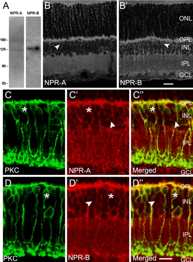
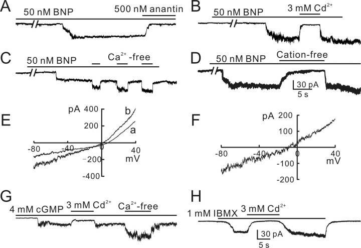
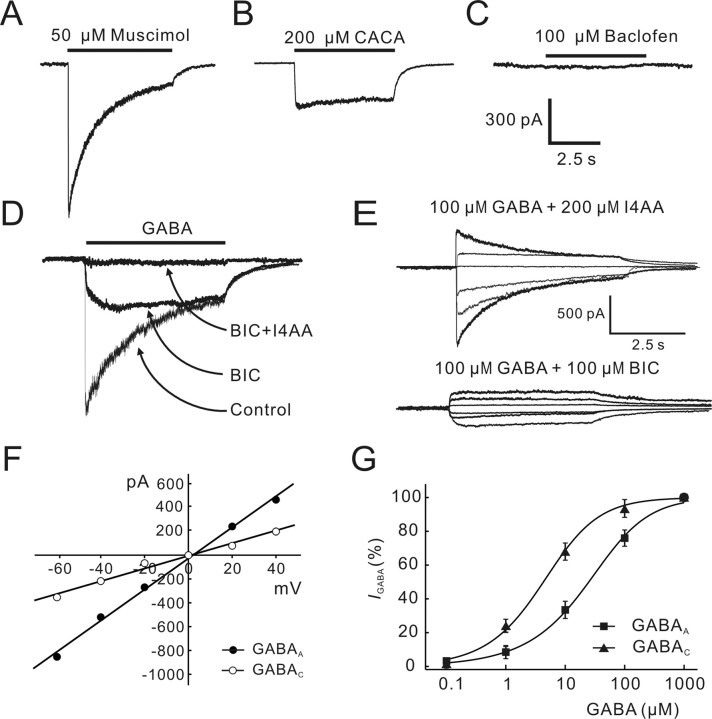
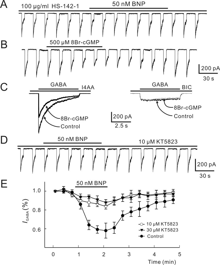
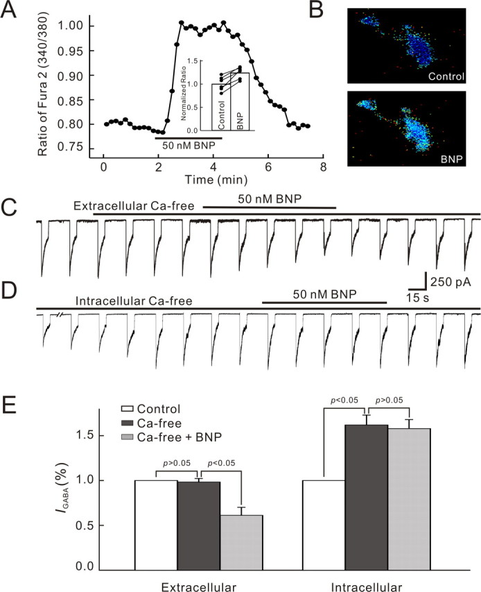
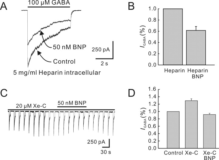
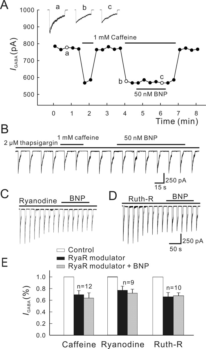
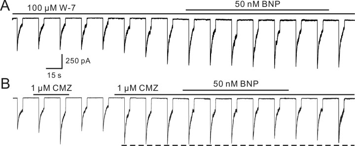
Natriuretic peptides (NPs) may work as neuromodulators through their associated receptors [NP receptors (NPRs)]. By immunocytochemistry, we showed that NPR-A and NPR-B were expressed abundantly on both ON-type and OFF-type bipolar cells (BCs) in rat retina, including the dendrites, somata, and axon terminals. Whole-cell recordings made from isolated ON-type BCs further showed that brain natriuretic peptide (BNP) suppressed GABAA receptor-, but not GABAC receptor-, mediated currents of the BCs, which was blocked by the NPR-A antagonist anantin. The NPR-C agonist c-ANF [des(Gln18, Ser19, Gln20, Leu21, Gly22)ANF(4-23)-NH2] did not suppress GABAA currents. The BNP effect on GABAA currents was abolished with preincubation with the pGC-A/B antagonist HS-142-1 but mimicked by application of 8-bromoguanosine-3',5'-cyclomonophosphate. These results suggest that elevated levels of intracellular cGMP caused by activation of NPR-A may mediate the BNP effect. Internal infusion of the cGMP-dependent protein kinase G (PKG) inhibitor KT5823 essentially blocked the BNP-induced reduction of GABAA currents. Moreover, calcium imaging showed that BNP caused a significant elevation of intracellular calcium that could be caused by increased calcium release from intracellular stores by PKG. The BNP effect was blocked by the ryanodine receptor modulators caffeine, ryanodine, and ruthenium red but not by the IP3 receptor antagonists heparin and xestospongin-C. Furthermore, the BNP effect was abolished after application of the blocker of endoplasmic reticulum Ca2+-ATPase thapsigargin and greatly reduced by the calmodulin inhibitors W-7 and calmidazolium. We therefore conclude that the increased calcium release from ryanodine-sensitive calcium stores by BNP may be responsible for the BNP-caused GABAA response suppression in ON-type BCs through stimulating calmodulin.


Figures

Figure 1.
Figure 1.
Immunolabeling of rat BCs with the anti-NP receptors antibodies. A, Western blots of whole rat retina extract using antibodies against NPR-A and NPR-B. In addition to a band at ∼125 kDa, corresponding to the size of the native rat NPR-A, the antibody to NPR-A revealed another band at ∼180 kDa, which might be attributable to glycosylation of the protein. The antibody to NPR-B revealed a single band at the corresponding molecular weight of 130 kDa. B, B′, Confocal laser microphotographs of the vertical sections of rat retinas, immunolabeled by the anti-NPR-A (B) and anti-NPR-B (B′) antibodies, respectively. The expression patterns of NPR-A and NPR-B are quite similar. Strong NPR-A and NPR-B immunoreactivities are clearly seen in both OPL and IPL. Note that lots of neurons in the INL are also NPR-A and NPR-B positive, some of which, located in the distal part of the layer (arrowheads), might be BCs. C-C″, D-D″, Double immunofluorescence labeling with the antibodies against NPR-A/B and PKC, a marker of ON-type BCs, respectively. C and C′ are the confocal micrographs of the vertical section of the retina, showing labeling for PKC (green) and NPR-A (red), respectively, and C″ is the merged image. PKC-positive ON-type BCs are characterized by a long axon terminating in sublamina b and an enlarged terminal bulb. Note that NPR-A is extensively expressed on the dendrites, somata, axons, and characteristic terminals of almost all the PKC-positive BCs. One of PKC-positive BCs is indicated by asterisks, and the labeling is rather strong on the cytomembrane. Somata of some cells in the INL, which are not labeled by PKC, are also NPR-A positive (arrowheads). These cells may be of OFF type. The expression profile of NPR-B is quite similar (D, D′). All parts of PKC-positive BCs, including the somata, dendrites, and axon terminals, are extensively labeled (asterisks). Again, some PKC-negative BCs in the distal part of INL are also positive to NPR-B (arrowheads). All of the micrographs were obtained by single optical sectioning at intervals of 1.0 μm. ONL, Outer nuclear layer. Scale bars, 10 μm.
Figure 2.
Figure 2.
Double immunofluorescence labeling of isolated rat bipolar cells. A-A″, Double labeling of an isolated BC with the antibodies against PKC and NPR-A. The cell that is PKC positive (A) is characterized by a long axon and an enlarged terminal bulb. The dendrites, axon terminal, and soma of the cell are all strongly labeled by NPR-A (A′). A″ is the merged image of A and A′. Note that the labeling is concentrated on the membrane. B-B″, Double labeling of another BC with similar morphology by the antibodies against PKC and NPR-B. The expression profile of NPR-B is quite similar. A0 and B0 are the Nomarski images of the two cells. C, D, Two PKC-negative BCs are also labeled by NPR-A (C) and NPR-B (D). PKC-negative BCs commonly exhibit distinct morphology, with much shorter axons. All parts of these cells are NPR positive. C′ and D′ are the Nomarski images of the cells shown in C and D, respectively. Scale bars, 10 μm.
Figure 3.
Figure 3.
BNP-induced inward currents in rat bipolar cells. A, BNP (50 nm) induced a sustained inward current from an isolated rat BC. The current emerged with a delay of ∼1.5 min and slowly rose to a steady level of 63 pA, and it was completely blocked by 500 nm anantin. B, BNP current was completely blocked by 3 mm Cd2+ applied externally in a reversible way. C, BNP current was potentiated by repetitive application of Ca2+-free bath solution with 10 mm EGTA added. D, BNP current was reversibly abolished when all external cations were replaced by choline (cation-free). The data shown in A-D were obtained in different BCs. E, I-V curves of a BC were obtained in the absence (a) and in the presence of 50 nm BNP (b) by presenting voltage ramp of 400 ms from -80 to 40 mV. F, The I-V curve of the BNP current, obtained by subtracting the data in curve a from the data in curve b, yielding a reversal potential of about -8.2 mV. G, Internal infusion of 4 mm cGMP induced a sustained inward current, which could be also blocked by 3 mm Cd2+ but potentiated by Ca2+-free extracellular solution. H, Perfusion of 1 mm IBMX also induced a sustained inward current from the cell, which could be blocked by 3 mm Cd2+. All the cells were voltage clamped at -60 mV, unless otherwise specified. Note the increased noise level in the currents induced by BNP, cGMP, and IBMX.
Figure 4.
Figure 4.
GABA-induced currents in isolated rat bipolar cells. A, Muscimol (50 μm) induced a current that desensitized to a steady level with a time constant of 1.24 s. B, CACA (200 μm) induced a rather sustained current. C, Baclofen (100 μm) did not induce any discernable current. The recordings shown in A-C were all made from the same cell. D, Current response of a BC to 100 μm GABA was partially suppressed by 100 μm BIC. Coapplication of 100 μm BIC and 200 μm I4AA completely blocked the current. E, BIC-sensitive GABAA currents (top) and I4AA-sensitive GABAC currents (bottom) recorded at different membrane potentials (-60, -40, -20, 0, 20, and 40 mV) from two BCs. F, I-V curves obtained using the data shown in E, yielding a reversal potential of 3.2 mV for the GABAA current (filled circles) and 1.3 mV for the GABAC current (open circles). G, Dose-response relationships of GABAA (filled squares) and GABAC (filled triangles) currents. GABAA currents were recorded in the presence of 200 μm I4AA, whereas GABAC currents were recorded in the presence of 100 μm BIC. EC50 of the GABAA current is 51.3 ± 6.2 μm (n = 6), and that of the GABAC current is 6.0 ± 0.6 μm (n = 4). The data for each cell were normalized by the maximum response of that cell to 1 mm GABA, and the normalized data were then averaged. The curves were drawn according to the following equation: I/Imax = 1/[1 + (EC50/[GABA])n]. Error bars represent SEM.
Figure 5.
Figure 5.
BNP suppresses GABAA, but not GABAC, current. GABA (100 μm) was repetitively applied for 5 s at intervals of 15 s. A, The GABA current was reduced when 50 nm BNP was applied. In this cell, 50 nm BNP did not induce a discernable inward current. B, BNP (50 nm) induced a sustained inward current in this cell. The GABA current was reduced by 50 nm BNP to a similar extent. Note that the full reduction of the GABA current was seen before the onset of the inward current. C, Comparison of BNP-caused reduction of GABA currents of BCs with or without BNP-induced inward currents. The two sets of data, which are represented as percentages of control in Ringer's, are not significantly different. IcGMP, cGMP-gated current. D, BNP (50 nm) induced an inward current in this cell and reduced the response of the cell to 100 μm GABA delivered for 5 s at intervals of 30 s. When the BNP-induced current was completely suppressed by cation-free solution, the reduction of the GABA current was almost the same to that observed in the presence of the BNP-induced current. GABA current excerpted at different times (a, b) are shown in the right panel at a faster time scale and a larger current scale. E, Anantin (500 nm) completely blocked the BNP-induced reduction of the GABA current of this cell. F, Application of 50 nm c-ANF did not change the GABA current. G, GABAA receptor-mediated current induced in a BC by coapplication of 100 μm GABA and 200 μm I4AA was reversibly reduced by 50 nm BNP. GABAC receptor-mediated current induced in a different cell by coapplication of 100 μm GABA and 100 μm BIC was not changed by 50 nm BNP. H, Averaged time courses of the effects of 50 nm BNP on GABAA and GABAC currents and timing of BNP application is indicated by the bar at the top. GABAA currents (filled circles) were reduced, whereas no significant reduction was found for GABAC currents (open circles). Error bars represent SEM.
Figure 6.
Figure 6.
Involvement of cGMP/PKG pathway in BNP-induced reduction of GABA currents. A, Internal infusion of HS-142-1 (100 μg/ml) for 5 min blocked the BNP-induced reduction of the GABA current. B, 8Br-cGMP (500 μm) applied extracellularly reversibly reduced the GABA response. Again, 100 μm GABA was repetitively delivered to the cell for 5 s at internals of 15 s. C, 8Br-cGMP (500 μm) reduced the GABAA current obtained by coapplication of GABA (100 μm) and I4AA (200 μm) (left) but did not change the GABAC current obtained in the same cell by coapplication of GABA (100 μm) and BIC (100 μm) (right). D, Internal infusion of KT5823 (10 μm) significantly attenuated the BNP-induced reduction of the GABA current. E, Changes in GABA currents caused by 50 nm BNP are plotted as a function of time during and after internal infusion of KT5823 of 10 μm (open circles) and 30 μm (filled triangles). Changes in GABA currents by 50 nm BNP without internal infusion of KT5823 (control) is presented for comparison (filled circles). Note that KT5823 did not completely block the BNP effect, and the data obtained with KT5823 of 10 or 30 μm are not very different. Error bars represent SEM.
Figure 7.
Figure 7.
Involvement of Ca2+ in BNP-induced reduction of GABA currents of bipolar cells. A, Continuous recording of intracellular calcium concentration given by the ratio of fura-2 AM fluorescence at 340 and 380 nm (340/380) from a BC using a fluorescence microscope equipped with a CCD camera. BNP (50 nm) was applied for ∼2 min, which remarkably increased [Ca2+]i in a reversible way. In the bar chart, shown in the inset, the BNP (50 nm)-caused changes in [Ca2+]i obtained in six cells are depicted. The average steady value obtained during BNP application in each cell was normalized to the value in normal Ringer's (control), and the normalized data were then averaged. Data obtained in individual cells are represented by solid lines. B, CCD pictures showing the change in [Ca2+]i caused by 50 nm BNP in a BC. Note that [Ca2+]i was clearly elevated in both soma and axon terminal. C, Ca2+-free extracellular solution (0 mm Ca2+ with 10 mm EGTA added) did not change the GABA current, and 50 nm BNP still suppressed the current in Ca2+-free extracellular solution. D, Internal infusion of Ca2+-free solution (0 mm Ca2+ with 10 mm BAPTA added) enhanced the GABA current and BNP failed to suppress the GABA current in Ca2+-free intracellular solution. E, Bar chart summarizing the effects of Ca2+-free extracellular solution and Ca2+-free intracellular solution on GABA currents and those of 50 nm BNP on GABA currents in these two solutions. Error bars represent SEM.
Figure 8.
Figure 8.
Effects of blockade of IP3-sensitive pathway on BNP-induced reduction of GABA currents. A, After internal infusion of 5 mg/ml heparin for 5 min, 50 nm BNP still reduced the GABA current. B, Bar chart showing that 50 nm BNP reduced the GABA current during internal infusion of heparin. C, Internal infusion of 20 μm xestospongin-C (Xe-C) increased the GABA current and did not block the BNP (50 nm)-induced reduction. D, Bar chart showing that internal infusion of xestospongin-C (Xe-C) increased the GABA current, compared with the initial value recorded just after membrane rupture. Application of 50 nm BNP induced a reduction of the GABA current in the presence of 20 μm xestospongin-C. Error bars represent SEM.
Figure 9.
Figure 9.
Effects of blockade of ryanodine-sensitive pathway on BNP-induced reduction of GABA currents. A, Caffeine (1 mm) greatly reduced the GABA current. In the presence of 1 mm caffeine, 50 nm BNP did not change the GABA current. For this experiment, GABA currents were induced every 15 s, and the current peaks were measured. Responses recorded at the times indicated by a, b, and c are shown in the top. B, Caffeine (1 mm) only slightly reduced the GABA current when the cell was preincubated with 2 μm thapsigargin, and 50 nm BNP failed to suppress the current in the presence of thapsigargin. C, Ryanodine (100 μm) caused a decrease of the GABA current, and 50 nm BNP did not change the GABA current during the internal infusion of ryanodine. D, Internal infusion of 20 μm ruthenium red (Ruth-R) showed similar effects. E, Bar chart summarizing the effects of the above treatments on GABA currents. Note that no changes in GABA currents after application of 50 nm BNP were observed in the presence of caffeine, ryanodine, or ruthenium red (Ruth-R). RyaR, Ryanodine receptor. Error bars represent SEM.
Figure 10.
Figure 10.
Involvement of calmodulin in BNP-induced reduction of GABA currents. A, Internal infusion of W-7 (100 μm) increased the GABA current and BNP (50 nm) no longer suppressed the GABA current in the presence of W-7. B, CMZ (1 μm) applied in external solution reversibly increased the GABA current. Note that 50 nm BNP-induced reduction of the GABA current was mainly attenuated, but was not completely blocked by, CMZ.

Similar articles

Cited by

References

    1. Aguayo LG, Espinoza F, Kunos G, Satin LS (1998) Effects of intracellular calcium on GABAA receptors in mouse cortical neurons. Pflügers Arch 435: 382-387. - PubMed
    1. Ahmad I, Leinders-Zufall T, Kocsis JD, Shepherd GM, Zufall F, Barnstable CJ (1994) Retinal ganglion cells express a cGMP-gated cation conductance activatable by nitric oxide donors. Neuron 12: 155-165. - PubMed
    1. Akopian A, Gabriel R, Witkovsky P (1998) Calcium released from intracellular stores inhibits GABAA-mediated currents in ganglion cells of the turtle retina. J Neurophysiol 80: 1105-1115. - PubMed
    1. Becchetti A, Roncaglia P (2000) Cyclic nucleotide-gated channels: intra- and extracellular accessibility to Cd2+ of substituted cysteine residues within the P-loop. Pflügers Arch 440: 556-565. - PubMed
    1. Bers DM, Patton CW, Nuccitelli R (1994) A practical guide to the preparation of Ca2+ buffers. Methods Cell Biol 40: 3-29. - PubMed

Publication types

MeSH terms

Substances

LinkOut - more resources