Comparative Study
. 2006 Jan 11;26(2):708-15.
doi: 10.1523/JNEUROSCI.4409-05.2006.

NMDA receptors in layer 4 spiny stellate cells of the mouse barrel cortex contain the NR2C subunit

Affiliations
Comparative Study

NMDA receptors in layer 4 spiny stellate cells of the mouse barrel cortex contain the NR2C subunit

Alexander M Binshtok et al. J Neurosci. .

Abstract

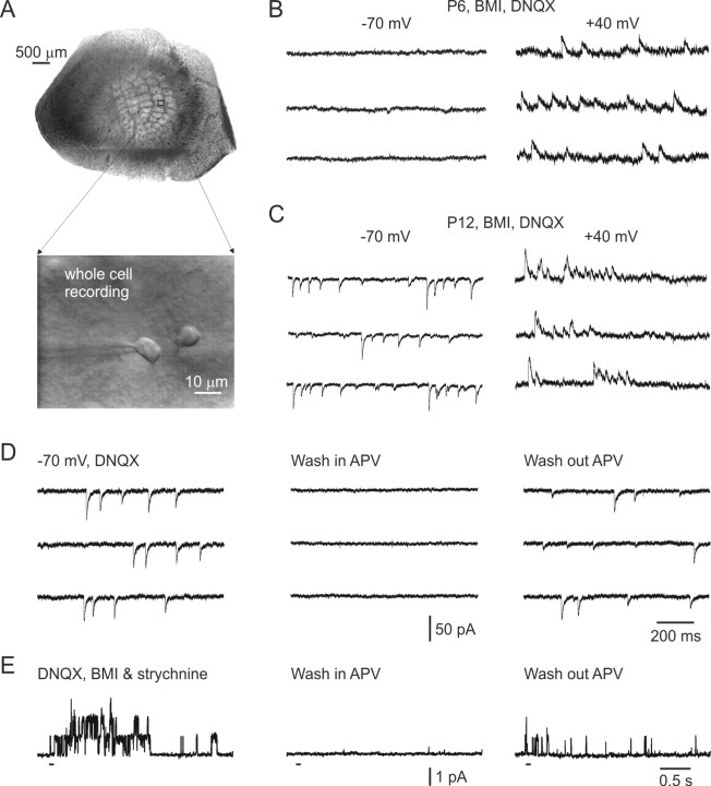
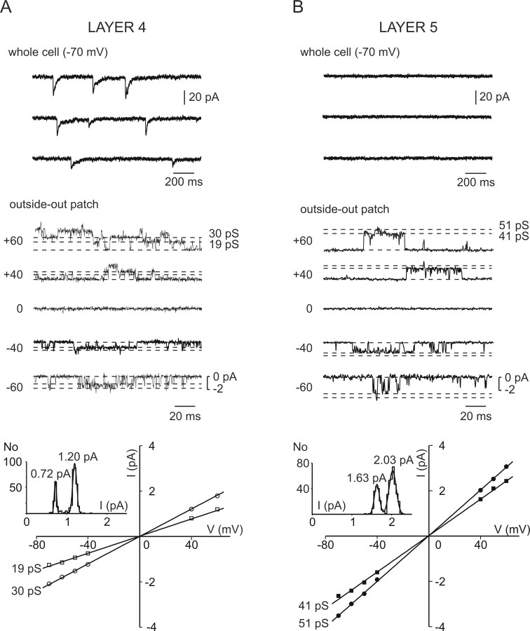
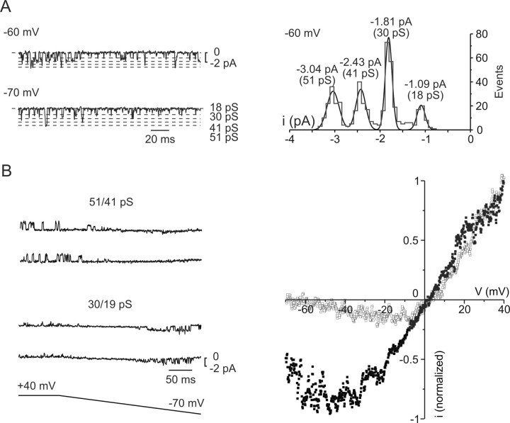
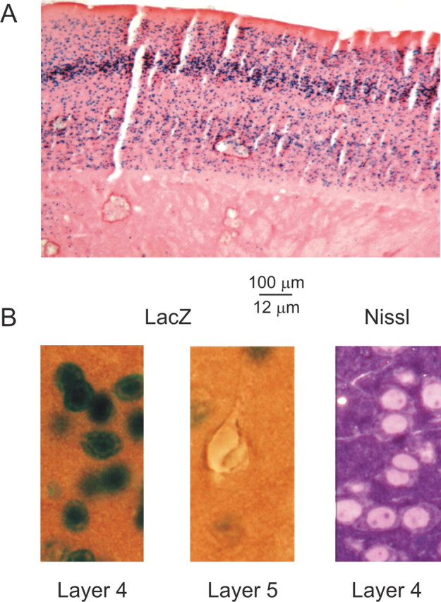
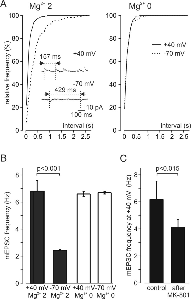
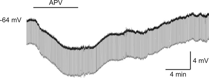
In layer 4 of the somatosensory cortex, the glutamatergic synapses that interconnect spiny stellate (SpS) neurons, which are the major targets of thalamocortical input, differ from most other neocortical excitatory synapses in that they have an extremely large NMDA receptor (NMDAR)-mediated component that is relatively insensitive to voltage-dependent Mg2+ blockade. We now report that this unique feature of the NMDA response reflects the distinctive subunit composition of the underlying receptors. We studied NMDAR-mediated miniature EPSCs (mEPSCs) and NMDA channel currents in tangential brain slices of mouse barrel cortex, which exclusively contain layer 4. NMDAR-mediated mEPSCs in SpS neurons were prominent at negative membrane potentials, and NMDA channels in outside-out patches excised from the somata of the same neurons had relatively low conductance and reduced susceptibility to Mg2+ block. These are characteristic features of heteromeric NMDAR assemblies that contain the NR2C subunit. Some patches also contained NMDA channels with higher conductance and a greater sensitivity to Mg2+. In the neocortex of transgenic mice in which a beta-galactosidase (lacZ) indicator gene was controlled by the NR2C promoter, the lacZ indicator was densely expressed in layer 4. In current-clamp recordings, blockade of NMDARs caused hyperpolarization and an increase in apparent input resistance. Our data demonstrate that the SpS neurons of layer 4 functionally express NR2C subunits; this is the likely explanation for their ability to generate large NMDAR-mediated EPSPs that are effective at resting potential, without previous depolarization.


Figures

Figure 1.
Figure 1.
In layer 4 of the mouse barrel cortex, NMDAR-mediated EPSCs at voltages near the resting potential appear after the first postnatal week. A, Top, Unstained 400-μm-thick tangential slice from a P20 mouse, containing layer 4 of the posteromedial barrel subfield, visualized at low magnification. Bottom, Whole-cell recording from identified layer 4 SpS neuron at higher magnification. B, At P6, whole-cell recording from a layer 4 SpS cell does not show any NMDAR-mediated activity at -70 mV, whereas activity is prominent when the membrane is depolarized to +40 mV. C, At P12, under the same experimental conditions, sEPSCs were prominent at both membrane potentials. D, Representative traces from a P22 SpS neuron demonstrate that, whereas inhibitory transmission was intact (no BMI was added into extracellular solution), APV-sensitive NMDAR-mediated sEPSCs were prominent at -70 mV. The recordings were performed with a CsGlu-containing electrode at -70 mV (EIPSCs). E, Brief pressure application (1-5 ms; 30 psi) of glutamate (0.2 mm) and glycine (0.1 mm) onto an outside-out patch (from the slice in A) elicited a long-lasting barrage of channel activity, which was completely and reversibly blocked by 50 μm APV. In this and all subsequent figures, aCSF contained 10 μm BMI and 20 μm DNQX.
Figure 2.
Figure 2.
Conductance properties of NMDA channels in layer 4 SpS neurons differ from those in layer 5 pyramidal neurons. A, Whole-cell and outside-out recordings from a layer 4 cell. Top, Prolonged NMDAR-mediated mEPSCs in a neuron held at -70 mV. Middle, Channel recordings from an outside-out patch excised from the same neuron and held at different voltages during exposure to a combination of 0.2 mm glutamate and 0.1 mm glycine. Dashed lines indicate the two conductance states and the closed state. Bottom, Current-voltage relationship obtained by measuring the amplitudes of 200-1000 well resolved openings for each membrane potential. The data are drawn from the patch shown in the middle panel. Linear regression analysis reveals slope conductances of 19 and 30 pS and a reversal potential of 0.2 mV. Inset, Amplitude distribution and fit with the sum of two Gaussians at +40 mV. B, Top, Under the same experimental conditions, the was no NMDAR-mediated activity in a layer 5 neuron at -70 mV. Middle, NMDA channel currents elicited by glutamate/glycine application to the outside-out patch, which was excised from the same neuron. Bottom, Current-voltage relationship and linear regression analysis provided estimated slope conductances of 41 and 51 pS and a reversal potential of 0.1 mV. Inset, Amplitude distribution and fit with the sum of two Gaussians at +40 mV.
Figure 3.
Figure 3.
Low-conductance NMDA channels in SpS neurons are less susceptible to Mg2+ blockade at negative membrane potentials. A, Left, Channel records from an outside-out patch that contains both high- and low-conductance type channels. Right, Amplitude distribution and fit with the sum of four Gaussians for voltage of -60 mV. B, Channel activities during voltage ramps were recorded, and traces containing only one kind of opening (left) were separated, and the currents were averaged to produce an instantaneous current-voltage relationship (right). Note that the inward current through 30/19 pS channel is significantly larger (closed squares) than that through 51/41 pS channel (open squares) over a wide voltage range (-10 to -70 mV).
Figure 4.
Figure 4.
Layer 4 neurons express the NR2C subunit of NMDAR. A, Horizontal section from the S1 region of a transgenic mouse that expressed a lacZ indicator gene controlled by the NR2C promoter region. Note that lacZ expression, while sparsely scattered throughout the layers, is particularly dense in layer 4. B, Somatic morphology of lacZ-expressing neurons in layer 4 (left) is very similar to that of SpS neurons, as shown in a 1 μm Nissl-stained section from a layer 4 brain slice previously used in a physiological experiment (right). lacZ is not expressed in layer 5 pyramidal neurons (middle).
Figure 5.
Figure 5.
Nonhomogeneous distribution of subtypes of NMDARs among individual synapses, as revealed by voltage dependence of mEPSCs. A, Left, Cumulative distribution of time intervals between NMDAR-mediated mEPSCs recorded at -70 mV (dashed line) and +40 mV (continuous line) in the presence of 2 mm Mg2+. Note that at -70 mV, the intervals between detectable mEPSCs are significantly longer. Inset, Sample current traces at -70 and +40 mV. Right, After Mg2+ wash out, mEPSC frequency at -70 mV increased, such that cumulative distribution of intervals between NMDAR-mediated mEPSCs at the two potentials did not differ. B, The voltage dependence of frequency of NMDAR-mediated mEPSCs. The bar graphs show mean frequency at -70 and +40 mV recorded in the presence of 2 mm Mg2+ (filled bars) and in Mg2+-free solution (open bars). C, In a different set of experiments, after 3 h of incubation in the presence of the open-channel NMDAR blocker MK-801 (40 μm), mEPSCs were completely blocked at -70 mV (data not shown), and mEPSC frequency at +40 mV was significantly decreased.
Figure 6.
Figure 6.
Tonic activation of NMDAR contributes to the resting membrane potential of SpS neurons. Current-clamp recording with a K gluconate-based, QX-314-containing pipette solution. To enhance glutamate release in the slice, extracellular K+ was increased to 5 mm, and Ca2+ was decreased to 1.2 mm. Note that the addition of 50 μm APV caused this neuron to hyperpolarize by 6 mV and its apparent input resistance, as revealed by voltage deflections during negative current pulses (20 pA, 1 s, 0.2 Hz), to increase by ∼26%.

Similar articles

Cited by

References

    1. Beattie EC, Carroll RC, Yu X, Morishita W, Yasuda H, von Zastrow M, Malenka RC (2000) Regulation of AMPA receptor endocytosis by a signaling mechanism shared with LTD. Nat Neurosci 3: 1291-1300. - PubMed
    1. Bliss TV, Collingridge GL (1993) A synaptic model of memory: long-term potentiation in the hippocampus. Nature 361: 31-39. - PubMed
    1. Brusa R, Zimmermann F, Koh DS, Feldmeyer D, Gass P, Seeburg PH, Sprengel R (1995) Early-onset epilepsy and postnatal lethality associated with an editing-deficient GluR-B allele in mice. Science 270: 1677-1680. - PubMed
    1. Carson F (1990) Histotechnology: a self-instructional text, Ed 1. Chicago: ASCP.
    1. Clark BA, Farrant M, Cull-Candy SG (1997) A direct comparison of the single-channel properties of synaptic and extrasynaptic NMDA receptors. J Neurosci 17: 107-116. - PMC - PubMed

Publication types

MeSH terms

LinkOut - more resources