. 2006 May 11;25(20):2890-900.
doi: 10.1038/sj.onc.1209308.

Erythroid progenitor renewal versus differentiation: genetic evidence for cell autonomous, essential functions of EpoR, Stat5 and the GR

Affiliations

Erythroid progenitor renewal versus differentiation: genetic evidence for cell autonomous, essential functions of EpoR, Stat5 and the GR

H Dolznig et al. Oncogene. .

Abstract

The balance between hematopoietic progenitor commitment and self-renewal versus differentiation is controlled by various transcriptional regulators cooperating with cytokine receptors. Disruption of this balance is increasingly recognized as important in the development of leukemia, by causing enhanced renewal and differentiation arrest. We studied regulation of renewal versus differentiation in primary murine erythroid progenitors that require cooperation of erythropoietin receptor (EpoR), the receptor tyrosine kinase c-Kit and a transcriptional regulator (glucocorticoid receptor; GR) for sustained renewal. However, mice defective for GR- (GR(dim/dim)), EpoR- (EpoR(H)) or STAT5ab function (Stat5ab(-/-)) show no severe erythropoiesis defects in vivo. Using primary erythroblast cultures from these mutants, we present genetic evidence that functional GR, EpoR, and Stat5 are essential for erythroblast renewal in vitro. Cells from GR(dim/dim), EpoR(H), and Stat5ab(-/-) mice showed enhanced differentiation instead of renewal, causing accumulation of mature cells and gradual proliferation arrest. Stat5ab was additionally required for Epo-induced terminal differentiation: differentiating Stat5ab(-/-) erythroblasts underwent apoptosis instead of erythrocyte maturation, due to absent induction of the antiapoptotic protein Bcl-X(L). This defect could be fully rescued by exogenous Bcl-X(L). These data suggest that signaling molecules driving leukemic proliferation may also be essential for prolonged self-renewal of normal erythroid progenitors.


Figures

Figure 1
Figure 1
Pre-expansion of proliferation-defective GRdim/dim-erythroblasts as multipotent cells yields erythroblasts defective for renewal but not for differentiation. (a) Signal transduction pathways important in erythropoiesis. Signaling molecules ablated or mutated in cells from genetically modified mice used in this study are indicated in red. (b) E12.5 fetal liver cells from GRdim/dim (white squares), GR−/− (gray triangles) or wild-type mice (WT, black circles) were cultivated in ‘erythroid proliferation medium’ and cumulative cell numbers determined daily. At day 12, aliquots were subjected to cytocentrifugation, stained and quantified for cells of increasing maturity (pie diagrams, blue, yellow, red) and apoptotic cells (black) as described (Kolbus et al., 2002). Cells (≥300) were counted per slide and mean values ±s.d. calculated from at least three independent determinations. Percentages of immature/partially mature/mature/dead cells were 70±6/5±1/9±2/16±3 for wild-type cells and 31±4/27±5/26±4/16±3 for GRdim/dim erythroblasts. (c) Cells from wild-type mouse fetal livers were pre-expanded in ‘stem cell mix’ medium (gray area, ‘SCM’), switched to erythroid conditions (day 9, white area, ‘Ery’) and cumulative cell numbers determined. Insets, cytospins from multipotent progenitors (top left) and cells switched to the erythroid lineage (day17, bottom right). (d) Multipotent cells (top panels, day9) and erythroblasts derived thereof (bottom panels, day 17, see panel (c) were subjected to flow cytometry. Percentages of cells single- (left) or double positive (right) for immature (green bars), erythroid (red bars), myeloid (green bars), and lymphoid surface markers (blue bars) detected by respective antibodies are shown. (e) Fetal liver cells from GRdim/dim- (white symbols) and wild-type (WT) mice (black symbols) were expanded in SCM, switched to the erythroid lineage (day 0, not shown) and analysed for differentiation kinetics in serum-containing differentiation medium. Cumulative cell numbers (left panel; mean values ±s.d., n=3), cell size (middle panel) and hemoglobin content (right panel) were determined at the times indicated.
Figure 2
Figure 2
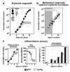
Erythroblasts expressing cytoplasmically truncated EpoRH: impaired renewal but normal differentiation. (a) Fetal liver-derived cells from EpoRH- (white symbols) and WT-mice (black) were cultivated in serum-free ‘erythroid proliferation medium’ and cumulative cell numbers determined as in Figure 1. Inset, scheme of mutated EpoR's used. (b) Cells as in (a) were pre-expanded in SCM (gray area, ‘SCM’) and switched to erythroid proliferation medium after 6 days (black arrow, white area, ‘Ery’). (c) After expansion for another 6 days in (gray arrow in (b) cells were analysed for differentiation parameters as in Figure 1 (three separate experiments, representative data shown).
Figure 3
Figure 3
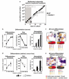
Erythroblasts from Stat5ab−/− mice: defects in renewal and terminal differentiation. (a) Fetal liver cells from Stat5ab−/− (white symbols) and wild-type mice (black) were expanded in SCM (gray area, ‘SCM’), switched to erythroid proliferation medium (white area, ‘Ery’), cultivated for another 6 days, and cumulative cell numbers were determined. (b, d) Stat5ab−/− and wild-type erythroid progenitors (day 12, gray arrow) analysed for differentiation parameters in both serum-containing (b) and defined, serum-free differentiation medium (d) as described in Figures 1 and 2 (error bars: s.d.s, n = 3). (c, e) Top panels: Cytospins from (b) and (d) subjected to neutral benzidine/histological staining after 48 h. Bottom: quantitation with respect to partially mature/mature/enucleated erythrocytes (percentages ±s.d.s, n = 3).
Figure 4
Figure 4
Bcl-XL upregulation in Stat5ab−/− erythroblasts is dependent on serum and not subject to direct transcriptional activation by Epo-driven Stat5 activation. (a) Primary Stat5ab−/− and wild-type erythroblasts were induced to differentiate in serum-containing (upper panels) or defined, serum-free differentiation medium (lower). Respective cell pellets (viable cells only) show hemoglobinization (red color, top of panels) and upregulation of Ter119 expression (data not shown, (Dolznig et al., 2001)). Lysates were analyzed for Bcl-XL protein (bottom) (b) Primary wild-type erythroblasts were induced to differentiate in serum-containing differentiation medium and analyzed for total Stat5ab protein, tyrosine-phosphorylated Stat5ab, and Bcl-XL at the times indicated. Stat5ab-DNA-binding activity was determined in EMSAs using extracts from the same preparations. (c) Upper panels: proliferating erythroblasts kept for 4 h without cytokines (no factor) and restimulated for 1 h with Epo (+ Epo), SCF (+ SCF) or both (+ Epo + SCF) were analysed by Northern blotting for bcl-XL mRNA and – as positive control – cis mRNA expression. Lower panels: primary mouse erythroblasts from wild-type (WT) or Stat5ab−/− fetal livers factor-depleted for 4 h, restimulated with Epo for 3 h and analysed for bcl-XL-mRNA (loading control: 18S ribosomal RNA) and protein expression.
Figure 5
Figure 5
Retrovirally expressed Bcl-XL rescues the differentiation but not the renewal defect of Stat5−/− erythroblasts. Freshly isolated wild type or Stat5ab−/− fetal liver cells were infected with MSCV-IRES-GFP or MSCV-Bcl-XL-IRES-GFP retroviral vectors (a) Cumulative cell numbers determined for Bcl-XL expressing wild-type or Stat5ab−/− erythroblasts expanded in ‘erythroid proliferation medium’. (b) Aliquots of proliferating cultures in (a) were subjected to cytospin analysis (arrow in a) for mature, partially mature, and immature cells as described for Figure 1 (top panels, viable cells only). The same cytospins were analysed for proportions of apoptotic/disintegrated cells and cells in mitosis, respectively (bottom) (c) The cell types shown in (a) and (b) were differentiated for 48 h in defined, serum-free medium lacking Epo. Cytospins (top) were quantified as in Figures 1 and 3 (percentages ±s.d.s, n = 3). (d) Aliquots from wild-type or Stat5ab−/− erythroblasts expressing empty vector (WT GFP, Stat5ab−/−) or exogenous Bcl-X (WT Bcl-XL, Stat5ab−/− Bcl-XL) were differentiated in fully defined medium plus Epo and analysed for hemoglobin content at the times indicated (error bars: s.d.s, n = 3).
Figure 6
Figure 6
Selective activation of Stat3 and Stat1 in proliferating and differentiating Stat5ab−/− erythroblasts. (a) Primary fetal liver cells from wild-type, Stat5ab+/−, and Stat5ab−/− mice were induced to differentiate in serum-free medium for 24 h and subjected to Western blot analysis for tyrosine-phosphorylated Stat5ab. (b) Primary wild-type- and Stat5ab−/− erythroblasts were induced to differentiate in defined serum-free medium for the times indicated and lysates were subjected to Western blot analysis for tyrosine phosphorylated Stat5, Stat3 and Stat1, as well as for total Stat5, Stat3, Stat1, and Bcl-XL protein. Loading control, eIF4E levels, *, nonspecific band.

Similar articles

Cited by

References

    1. Bauer A, Gandrillon O, Samarut J, Beug H. In: Hematopoiesis: a developmental approach. Zon L, editor. Oxford University Press; Oxford: 2001. pp. 368–390.
    1. Bauer A, Tronche F, Wessely O, Kellendonk C, Reichardt HM, Steinlein P, et al. Genes Dev. 1999;13:2996–3002. - PMC - PubMed
    1. Beug H, Bauer A, Dolznig H, von Lindern M, Lobmayer L, Mellitzer G, et al. Biochim Biophys Acta. 1996;1288:M35–M47. - PubMed
    1. Beug H, Dahl R, Steinlein P, Meyer S, Deiner E, Hayman MH. Oncogene. 1995;11:59–72. - PubMed
    1. Blau HM, Brazelton TR, Weimann JM. Cell. 2001;105:829–841. - PubMed

Publication types

MeSH terms