. 2006 May 4;25(19):2785-94.
doi: 10.1038/sj.onc.1209303.

N-(4-hydroxyphenyl)retinamide-induced apoptosis triggered by reactive oxygen species is mediated by activation of MAPKs in head and neck squamous carcinoma cells

Affiliations

N-(4-hydroxyphenyl)retinamide-induced apoptosis triggered by reactive oxygen species is mediated by activation of MAPKs in head and neck squamous carcinoma cells

H-J Kim et al. Oncogene. .

Abstract

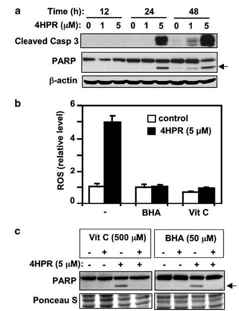
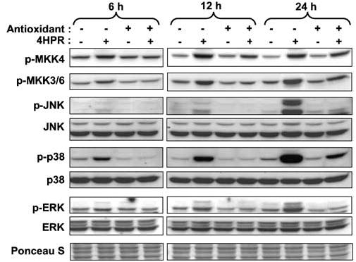
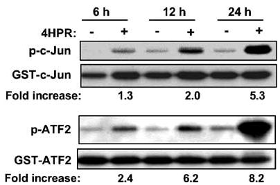
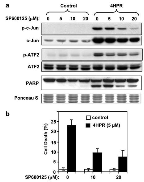
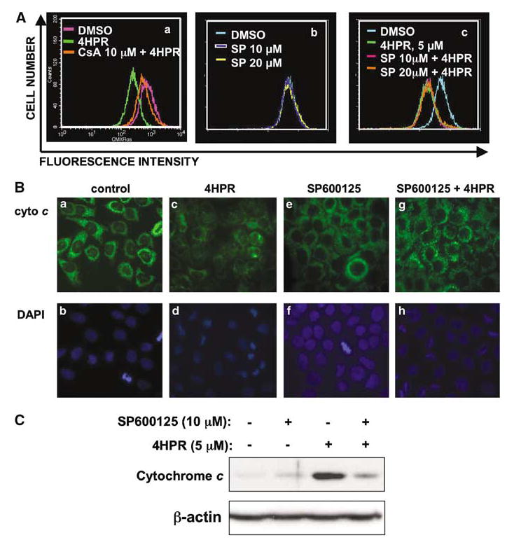
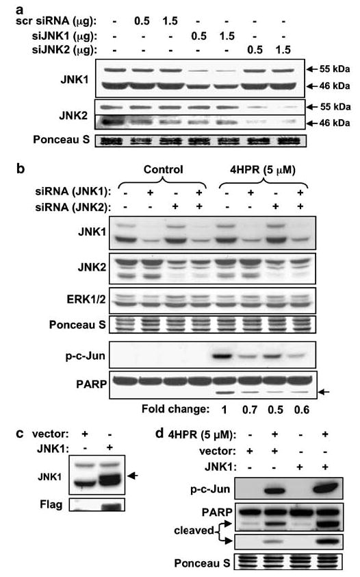
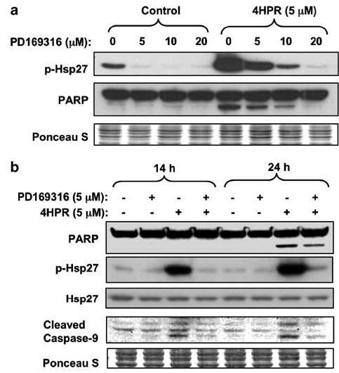
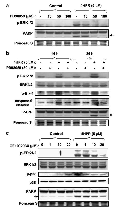
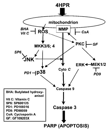
N-(4-hydroxyphenyl)retinamide (4HPR), a synthetic retinoid effective in cancer chemoprevention and therapy, is thought to act via apoptosis induction resulting from increased reactive oxygen species (ROS) generation. As ROS can activate MAP kinases and protein kinase C (PKC), we examined the role of such enzymes in 4HPR-induced apoptosis in HNSCC UMSCC22B cells. 4HPR increased ROS level within 1 h and induced activation of caspase 3 and PARP cleavage within 24 h. Activation of MKK3/6 and MKK4, JNK, p38 and ERK was detected between 6 and 12 h, increased up to 24 h and preceded apoptosis. 4HPR-induced activation of these kinases was abrogated by the antioxidants BHA and vitamin C. SP600125, a JNK inhibitor, suppressed 4HPR-induced c-Jun phosphorylation, cytochrome c release from mitochondria and apoptosis. Suppression of JNK1 and JNK2 using siRNA decreased, whereas overexpression of wild type-JNK1 enhanced 4HPR-induced apoptosis. PD169316, a p38, inhibitor suppressed phosphorylation of Hsp27 and apoptosis. PD98059, an MEK1/2 inhibitor, also suppressed ERK1/2 activation and apoptosis induced by 4HPR. Likewise, PKC inhibitor GF109203X suppressed ERK and p38 phosphorylation and PARP cleavage. These data indicate that 4HPR-induced apoptosis is triggered by ROS increase, leading to the activation of the mitogen-activated protein serine/threonine kinases JNK, p38, PKC and ERK, and subsequent apoptosis.


Figures

Figure 1
Figure 1
Induction of apoptosis by 4HPR and its suppression by antioxidants. (a) Time course of activation of caspase-3 and cleavage of PARP induced by 4HPR. The cells were treated with 4HPR (1 or 5 μm as indicated) or DMSO control for 12, 24, or 48 h. Total cell lysates were subjected to SDS–PAGE, followed by Western blotting with specific antibodies against cleaved caspase-3 and PARP, followed by anti-actin antibodies to ascertain equal loading. (b) Effects of antioxidants on ROS generation were determined after 60 min of incubation with 4HPR or 4HPR plus BHA or vitamin C using the DCF-DA assay. (c) Effect of antioxidants on 4HPR-induced PARP cleavage was determined by Western blotting analysis. Arrows point to the location of the cleaved PARP fragment. Ponceau S staining was used to compare loading in different lanes.
Figure 2
Figure 2
Effects of antioxidants (BHA and vitamin C) on 4HPR-induced activation of MKKs (MKK4 and MKK3/6) and their downstream substrates MAPKs (JNK, p38 and ERK). Cells were treated with 4HPR (5 μm) alone or with antioxidant (BHA (50 μm) for MKK4, JNK and ERK, and vitamin C (500 μm) for MKK3/6 and p38) for the indicated time periods. The cells were then harvested and total cell lysates were prepared and resolved on 10% SDS–PAGE and subjected to Western blotting analysis with antibodies against the different protein kinases.
Figure 3
Figure 3
Effects of 4HPR on JNK and p38 kinase activities. Cells were treated with DMSO or 4HPR (5 μm) for the indicated times. The cells were then harvested, solubilized and the lysates were used for JNK and p38 kinase assays with GST-c-Jun and GST-ATF-2 as substrates, respectively (see Materials and methods). Data from a representative experiment from among three independent experiments with similar findings are shown. The phosphorylation was quantitated by densitometry using the NIH image analysis program. ‘Fold increase’ is expressed relative to the control value obtained with DMSO-treated cells (−).
Figure 4
Figure 4
Effects of a JNK inhibitor on 4HPR-induced apoptosis. (a) Cells were pretreated with the indicated concentrations of SP600125 for 2 h. Then 4HPR (5 μm) was added to the culture medium and cells were incubated for an additional 24 h. Phosphorylation status of the JNK substrates c-Jun and ATF-2 was determined by Western blotting with phospho-specific antibodies. PARP cleavage was determined by re-probing the membranes using specific antibodies. (b) Inhibition of 4HPR-induced cell death by SP600125 was quantified by nuclear DAPI staining as described in ‘Materials and methods’. The data are the mean ± s.d. of triplicates.
Figure 5
Figure 5
Effect of JNK inhibitor on mitochondrial events induced by 4HPR. (A) Changes in mitochondrial membrane potential were monitored by using cell-permeable mitochondria-selective dye CMXRos as described in ‘Materials and methods’. (a) Cells were pretreated with cyclosporine A (CsA) for 1 h, followed by incubation either with DMSO or 4HPR for an additional 1 h. (b, c) Cells were pretreated with SP600125 for 2 h, followed by incubation with DMSO or 4HPR for an additional 1 h. (B) Subcellular localization of cytochrome c was determined by immunofluorescence. Cells were pretreated with DMSO or SP600125 (10 μm) for 2 h and subsequently incubated with 4HPR for 14 h. The cells were then subjected to immunofluorescent labeling to localize cytochrome c as described in ‘Materials and methods.’ (a, c, e, g) cytochrome c staining. (b, d, f, h) nuclear DAPI staining. (C) Detection of cytochrome c by Western blotting analysis. After cells were treated with either 4HPR alone or in combination with SP600125, cytosolic proteins were isolated and subjected to Western blot analysis using cytochrome c antibody. The membrane was probed with β-actin antibodies to compare loading in different lanes.
Figure 6
Figure 6
Demonstration of the role of JNK1 and JNK2 in 4HPR-induced cell death using loss- and gain-of-function approaches. (a) Effect of siRNA against JNK1 and JNK2 on endogenous JNK1 and JNK2 protein levels. Cells were transfected with siRNA against JNK1, JNK2 or scrambled siRNA. The protein levels of JNK1 and JNK2 were determined by Western blotting 1 day after transfection by using antibodies against JNK1 and JNK2. (b) Effects of siRNA against JNK1 and JNK2 on 4HPR-induced cell death. Cells transfected with siRNA against JNK1, JNK2 or combination of the two were treated with 4HPR for 24 h and total cell lysates were subjected to Western blot analysis using the antibodies indicated. (c) Cells were transiently transfected with either empty vector or wt-JNK1 expressing vector. The levels of endogenous and exogenously expressed JNK1 were determined by Western blotting 24 h after transfection. (d) Cells transfected with wt-JNK1 were treated with 4HPR for 24 h. Total cell lysates were analysed by Western analysis to determine the level of phospho-c-Jun and cleaved PARP.
Figure 7
Figure 7
Effects of a p38/MAPK inhibitor on 4HPR-induced apoptosis. (a) Cells were pretreated with PD169316 at different doses for 2 h, followed by incubation with 4HPR for an additional 24 h. The phosphorylation status of the Hsp27 was determined by Western blotting with phospho-specific antibody as an indication of p38 kinase activity. (b) Cells treated with 4HPR alone or in combination with PD169316 for the indicated time periods were analysed by Western blotting with indicated antibodies.
Figure 8
Figure 8
Effect of MEK1/2 inhibitor (PD98059) and PKC inhibitor (GF109203X) on 4HPR-induced apoptosis. (a) Cells were pretreated with PD98059 with the indicated concentrations for 2 h followed by incubation with 4HPR for additional 24 h. Phosphorylation status of ERK1/2 (a substrate of MEK1/2) was determined by Western blotting with phospho-specific antibody. (b) Cells were pretreated with 50 μm PD98059 for 2 h. 4HPR was then added to the culture medium and cells were incubated for additional 14 or 24 h. Phosphorylation status of the kinase target proteins was determined by Western blotting with phospho-specific antibodies against ERK and Elk-1. PARP cleavage, a marker of caspase-3 activation and apoptosis, and caspase-9 cleavage were determined by re-probing the membranes using specific antibodies. (c) Cells were pretreated with the indicated concentrations of GF109203X for 2 h, followed by incubation with 4HPR for additional 24 h. Phosphorylation status of ERK1/2 and p38 was determined by Western blotting with phospho-specific antibodies and antibodies against total ERK1/2 and p38. PARP cleavage was determined by re-probing the membranes using specific antibodies. The membranes were stained with Ponceau S to compare loading in different lanes.
Figure 9
Figure 9
Schematic representation of the different pathways shown in this report to be activated by 4HPR through ROS and lead to apoptosis in HNSCC cells. The use of antioxidants and various inhibitors of kinases indicates that 4HPR acts on the mitochondria to increase the production of ROS and alter the mitochondrial membrane potential. The increased membrane permeability facilitates the release of cytochrome c to the cytoplasm. The increase in ROS also activates PKC and MKKs. The activation of PKC further augments cytochrome c release, as well as activates p38 and MEK1/2. The activation of ERK appears to be associated with apoptosis induction. The activation of MKKs results in activation of JNK and p38, which could enhance apoptosis via the cytochrome c, caspase-9 and caspase-3 pathway.

Similar articles

Cited by

References

    1. Aikawa R, Komuro I, Yamazaki T, Zou Y, Kudoh S, Tanaka M, et al. J Clin Invest. 1997;100:1813–1821. - PMC - PubMed
    1. Asumendi A, Morales MC, Alvarez A, Arechaga J, Perez-Yarza G. Br J Cancer. 2002;86:1951–1956. - PMC - PubMed
    1. Bennett BL, Sasaki DT, Murray BW, O’Leary EC, Sakata ST, Xu W, et al. Proc Natl Acad Sci USA. 2001;98:13681–13686. - PMC - PubMed
    1. Bhatt NY, Kelley TW, Khramtsov VV, Wang Y, Lam GK, Clanton TL, et al. J Immunol. 2002;169:6427–6434. - PubMed
    1. Boya P, Morales MC, Gonzalez-Polo RA, Andreau K, Gourdier I, Perfettini JL, et al. Oncogene. 2003;22:6220–6230. - PubMed

Publication types

MeSH terms