. 2006 Jan 12;439(7073):220-4.
doi: 10.1038/nature04375.

Planar cell polarity signalling couples cell division and morphogenesis during neurulation

Affiliations

Planar cell polarity signalling couples cell division and morphogenesis during neurulation

Brian Ciruna et al. Nature. .

Abstract

Environmental and genetic aberrations lead to neural tube closure defects (NTDs) in 1 out of every 1,000 births. Mouse and frog models for these birth defects have indicated that Van Gogh-like 2 (Vangl2, also known as Strabismus) and other components of planar cell polarity (PCP) signalling might control neurulation by promoting the convergence of neural progenitors to the midline. Here we show a novel role for PCP signalling during neurulation in zebrafish. We demonstrate that non-canonical Wnt/PCP signalling polarizes neural progenitors along the anteroposterior axis. This polarity is transiently lost during cell division in the neural keel but is re-established as daughter cells reintegrate into the neuroepithelium. Loss of zebrafish Vangl2 (in trilobite mutants) abolishes the polarization of neural keel cells, disrupts re-intercalation of daughter cells into the neuroepithelium, and results in ectopic neural progenitor accumulations and NTDs. Remarkably, blocking cell division leads to rescue of trilobite neural tube morphogenesis despite persistent defects in convergence and extension. These results reveal a function for PCP signalling in coupling cell division and morphogenesis at neurulation and indicate a previously unrecognized mechanism that might underlie NTDs.


Figures

Figure 1
Figure 1
PCP signalling is required for zebrafish neural tube formation. (a-h) Confocal micrographs of transverse sections through rhodamine-phalloidin stained embryos, comparing WT and MZtri neural tube morphogenesis at 5-somite/neural plate (a,b), 10-somite/neural keel (c,d), 15-somite/neural rod (e,f), and 20-somite/neural tube (g,h) stages. The neural anlage has been outlined in all panels. (i-j) Sections through mGFP-injected 20-somite staged embryos showing ectopic cell accumulations within the developing neural tube of MZslb;MZppt embryos (i), and severe disorganization of the neural anlage in MZslb;MZppt embryos that were injected with 6ng of Wnt4 MO (j). (k-m) Rhodamine-phalloidin stained sections through 20-somite staged MZtri embryos cultured overnight in either 4% DMSO (k), or in the presence cell division inhibitors (l-m). Note rescue of the expanded floorplate (arrows in k-m) and neural tube morphogenesis defects upon blocking cell division. The extent of ectopic cellular accumulations in PCP signalling mutants has been highlighted (h-l). Scale bars, 50 μm.
Figure 2
Figure 2
Cell autonomy of PCP signalling within the neural keel. (a-b) Whole-mounts and transverse sections through the trunk of 24 hour post-fertilization WT (a) and MZtri (b) embryos injected with 100 pg of lefty mRNA. Convergence of the neural plate into a neural rod occurs normally in WT+lefty embryos, despite the absence of underlying mesendoderm (a'). This convergence is disrupted in MZtri+lefty mutants, which show neurulation defects in the absence of trunk mesoderm (b'). Brackets in a and b indicate the extent of trunk axial extension. Identical results were obtained using a different genetic combination: maternal-zygotic one-eyed-pinhead (MZoep) mutants were used to eliminate Nodal signalling, and PCP signalling was perturbed through diego mRNA injection (not shown). (c) mGFP-labelled MZtri cells were transplanted into MZoep host embryos at mid-blastula stages to generate MZtri->MZoep chimeric embryos. MZoep mutants lack Nodal signalling and do not form endoderm or trunk mesoderm lineages, therefore endoderm and trunk somites in chimeric embryos develop entirely from GFP-positive MZtri donor cells. (c') Transverse sections of 24 hpf MZtri->MZoep chimeras were counter-stained with rhodamine-phalloidin to visualize neural tube formation (outlined in c'). The absence of MZtri neurulation defects indicates a requirement for PCP signalling autonomous to the neuroectoderm. (d-g) Transverse sections through the trunk of WT and MZtri chimeric embryos at 24 hpf. (d) Bilateral distribution of mRFP-labelled WT cells (arrowheads) within the neural tube of an mGFP-labelled WT host. (e) Unilateral accumulation of mRFP-labelled MZtri cells within the neural tube of mGFP-labelled WT host embryos. (f-g) mRFP-labelled MZtri (f) or WT (g) cells transplanted into mGFP-labelled MZtri host embryos accumulate unilaterally within the MZtri neural anlage. Scale bars, 50 μm.
Figure 3
Figure 3
The cellular basis of MZtri neurulation defects. (a-d) Lineage tracing of WT (a,b) and MZtri (c,d) neural plate cells after red photo-conversion of the Kaede fluorophore. WT neural progenitors routinely crossed the midline into the contralateral side of the neural tube (b). MZtri neural progenitor cells never integrated into the contralateral neuroepithelium (d). Cells accumulating ectopically in the centre of the MZtri neural anlage also respected the midline (dashed lines). (e-m) Confocal micrographs from time series depicting cell division during WT (e-g) and MZtri (h-m) neurulation. Time is indicated as minutes preceding or following the completion of cytokinesis. The boundary of the neural keel has been highlighted, and the midline indicated. Cells in the WT neural keel round up their cell bodies and divide apically (e). Basal daughter cells remain connected to the basement membrane via a cellular process (arrow in e,f) and re-insert into the neuroepithelium (asterisk in g). Apical daughter cells (arrow head in f,g) lose contact within the basal membrane, adopt medial-lateral polarity, and intercalate across the midline of the neural keel. MZtri cells divide apically, and basal daughter cells behave as in WT (arrow in h,i,k,l, asterisk in j). MZtri apical daughter cells (arrowhead in i,j,l,m) do not intercalate into the contralateral neuroepithelium and remain in the place they were born. Scale bars, 50 μm.
Figure 4
Figure 4
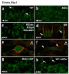
Anterior membrane localization of Gfp-Pk as a marker of planar polarity. Confocal images taken at the level of the anterior spinal cord, through the dorsal-ventral plane of the neural keel (a-c, e-h) or notochord (d) of 8- to 10-somite staged embryos. (a-c) Scatter labelling of Gfp-Pk in the neural keel of (a) a WT embryo, (b) an MZtri mutant, or (c) an MZslb;MZppt mutant injected with 6 ng of Wnt4 MO. (d) Scatter labelling of Gfp-Pk plus mRFP in WT notochord, demonstrating anterior membrane translocation of Gfp-Pk (arrows) in cells that undergo well-characterized CE movements in response to PCP signalling. (e-f) A Gfp-Pk plus mRFP labelled WT neural keel cell demonstrating transient loss of polarity markers during mitosis (e), but re-establishment of membrane-localized Gfp-Pk in both daughter cells (arrows in f). (g-h) Chimeric analysis of the autonomy of PCP signalling, using Gfp-Pk as a marker of planar polarity. (g) MZtri cells transplanted into WT host embryos do not localize Gfp-Pk to the membrane, as Vangl2 is required for Pk translocation. (h) WT cells transplanted into MZtri hosts show reduced membrane Gfp-Pk localization and abnormal polarity (arrows). “Ant” marks the anterior direction.

Comment in

References

    1. Copp AJ, Greene ND, Murdoch JN. The genetic basis of mammalian neurulation. Nat Rev Genet. 2003;4:784–93. - PubMed
    1. Greene ND, Gerrelli D, Van Straaten HW, Copp AJ. Abnormalities of floor plate, notochord and somite differentiation in the loop-tail (Lp) mouse: a model of severe neural tube defects. Mech Dev. 1998;73:59–72. - PubMed
    1. Kibar Z, et al. Ltap, a mammalian homolog of Drosophila Strabismus/Van Gogh, is altered in the mouse neural tube mutant Loop-tail. Nat Genet. 2001;28:251–5. - PubMed
    1. Murdoch JN, Doudney K, Paternotte C, Copp AJ, Stanier P. Severe neural tube defects in the loop-tail mouse result from mutation of Lpp1, a novel gene involved in floor plate specification. Hum Mol Genet. 2001;10:2593–601. - PubMed
    1. Keller R. Shaping the vertebrate body plan by polarized embryonic cell movements. Science. 2002;298:1950–4. - PubMed

Publication types