. 2006 Feb 8;25(3):585-95.
doi: 10.1038/sj.emboj.7600939. Epub 2006 Jan 12.

SMUG1 is able to excise uracil from immunoglobulin genes: insight into mutation versus repair

Affiliations

SMUG1 is able to excise uracil from immunoglobulin genes: insight into mutation versus repair

Javier M Di Noia et al. EMBO J. .

Abstract

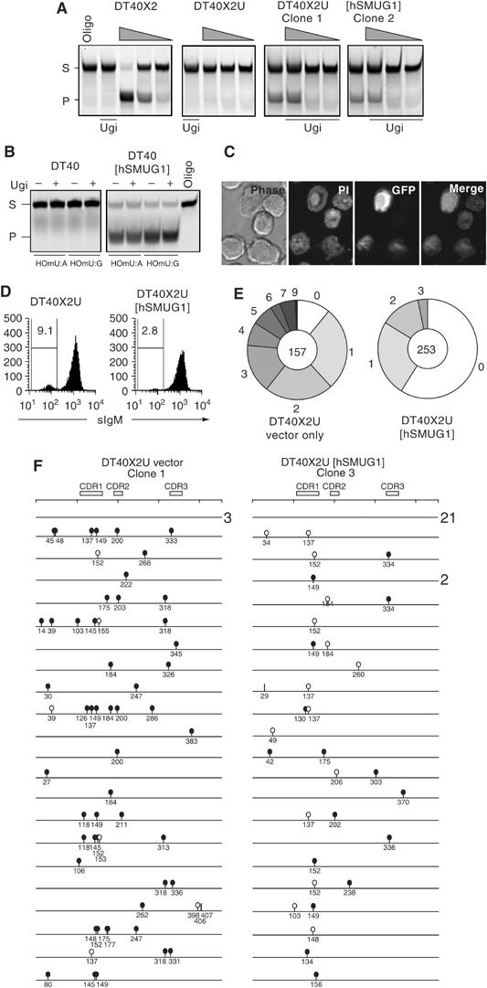
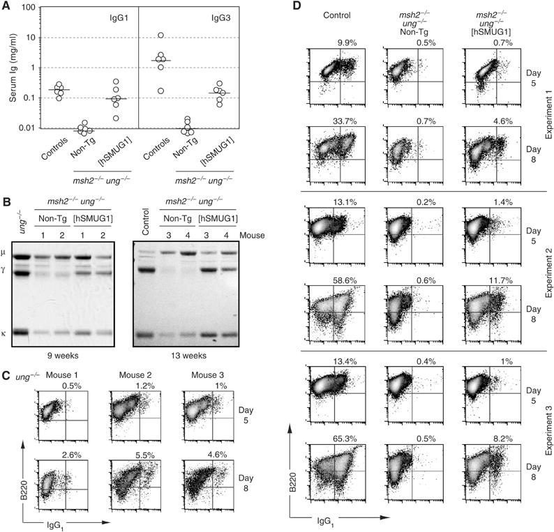
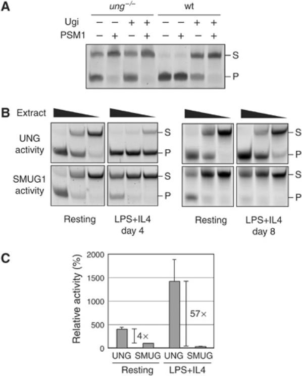
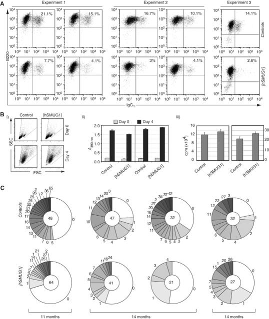
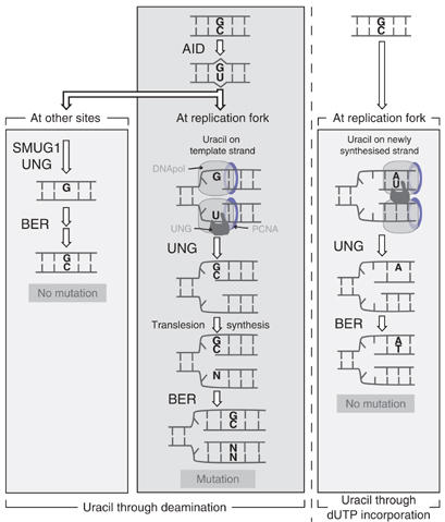
Mammals harbour multiple enzymes capable of excising uracil from DNA, although their distinct physiological roles remain uncertain. One of them (UNG) plays a critical role in antibody gene diversification, as UNG deficiency alone is sufficient to perturb the process. Here, we show this unique requirement for UNG does not reflect the fact that other glycosylases are unable to access the U:G lesion. SMUG1, if overexpressed, can partially substitute for UNG to assist antibody diversification as judged by its effect on somatic hypermutation patterns (in both DT40 B cells and mice) as well as a restoration of isotype switching in SMUG-transgenic msh2-/- ung-/- mice. However, SMUG1 plays little natural role in antibody diversification because (i) it is diminishingly expressed during B-cell activation and (ii) even if overexpressed, SMUG1 more appears to favour conventional repair of the uracil lesion than assist diversification. The distinction between UNG and overexpressed SMUG1 regarding the balance between antibody diversification and non-mutagenic repair of the U:G lesion could reflect the association of UNG (but not SMUG1) with sites of DNA replication.


Figures

Figure 1
Figure 1
Expression of hSMUG1 in DT40 alters the mutation spectrum and reduces mutation accumulation. (A) Comparison of uracil excision activity in extracts of XRCC2-deficient derivatives of DT40 (DT40X2) that express Ugi (DT40X2U) or Ugi together with human SMUG1 (DT40X2U[hSMUG1]). Uracil excision activity is reflected by cleavage of the fluorescently labelled 42-mer U-containing oligonucleotide substrate (S) into a 26-nucleotide product (P). The gel was loaded with samples containing either oligonucleotide substrate alone (oligo) or following incubation with serial five-fold dilutions of extract (wedges). Extracts were preincubated on ice for 20 min with Ugi where indicated. (B) Assay for 5-hydroxymethyl uracil (HOmU) excision activity in extracts of DT40 transfectants expressing hSMUG1 as compared to controls. Activity was measured using double-stranded HOmU-containing oligonucleotide substrates in which the HOmU residue was paired with either A or G: extracts were preincubated with exogenous Ugi where indicated. (C) Confocal images of DT40 cells expressing an hSMUG1-GFP fusion protein: cellular DNA was stained with propidium iodide (PI). (D) Flow cytometric analysis of surface IgM expression in one representative DT40X2U[hSMUG1] and one vector-only-transfected DT40X2U control clone that had been expanded for 10 weeks following single cell cloning. The proportions of sIgM negatives (%) are indicated, with the results from multiple clones summarised in Table II. (E) Pie chart depicting the proportion of IgVλ sequences bearing the indicated number of mutations. The data from four DT40X2U[hSMUG1] and three DT40X2U control clone have been separately pooled. The total number of sequences in each pool is indicated at the centre of the pie. (F) Schematic depiction of the nature of the identified IgVλ mutations. All the 43 sequences obtained from one representative DT40X2U[hSMUG1] clone are shown (22 mutated; 21 unmutated) together with the first 24 sequences (21 mutated; three unmutated) from a vector-only control clone. Numbers at the right indicate when the same sequence was found more than once. Transition mutations at C:G are indicated by black lollipops, transversion mutations at C:G by white lollipops and mutations at A:T pairs by vertical lines.
Figure 2
Figure 2
SMUG1 overexpression in msh2−/−ung−/− mice allows the accumulation of substantial levels of serum IgG. (A) Titres of IgG1 and IgG3 in sera of mice of different genotypes (aged 9–20 weeks) as determined by ELISA. The mice analysed were the products of crosses between msh2+/−ung−/− [hSMUG1] transgenic males and msh2+/−ung−/− non-transgenic females, with age-matched C57BL/6 mice providing controls. (B) Serum immunoglobulin profiles of four msh2−/−ung−/− control and four msh2−/−ung−/− [hSMUG1] transgenic mice analysed at the ages indicated. Representative examples of sera from age-matched normal and ung−/− mice are shown for comparison. Total serum immunoglobulin was precipitated using protein L-agarose, resolved by SDS–PAGE and stained with Coomassie blue. (C) Flow cytometric profiles of three independent experiments in which splenic B cells from UNG-deficient mice were cultured in the presence of LPS+IL4 for 5 or 8 days prior to staining for sIgG1 and CD45R(B220). Live cells were gated by staining with propidium iodide, and percentages of sIgG1+ B cells are indicated. (D) Flow cytometric profiles of three independent experiments in which splenic B cells from msh2−/−ung−/− [hSMUG1]-transgenic, msh2−/−ung−/− control or wild-type mice were cultured in the presence of LPS+IL4 for 5 or 8 days prior to staining for sIgG1 and CD45R(B220). Live cells were gated and percentages indicated as in panel C.
Figure 3
Figure 3
Comparison of SMUG1 and UNG uracil excision activity during in vitro B-cell activation. (A) Uracil excision activity in whole cell extracts of splenic B cells from either wild-type or ung−/− mice following activation for 4 days with LPS+IL4 was assayed using a 42-mer double-stranded oligonucleotide substrate. Where indicated, the extracts were incubated for 20 min on ice with the UNG inhibitor Ugi or with the PSM1 anti-SMUG1 antibody before adding the substrate. (B) Comparison of uracil excision activity in serial five-fold dilutions of whole cell extracts prepared from splenic B cells of a wild-type mouse after 0, 4 or 8 days of incubation with LPS+IL4. UNG activity was determined by performing the uracil excision assay in the presence of the PSM1 anti-SMUG1 antibody, whereas SMUG1 activity was determined by performing the assay in the presence of Ugi. (C) Quantitation of the relative levels of UNG and SMUG1 activity in splenic B cell extracts prepared after 0 or 4 days of incubation with LPS+IL4. The histogram is based on the results of four pairs of mice analysed as in panel B. UNG and SMUG1 activities were quantified by gel densitometry using the IQ software (Molecular Dynamics) and expressed as mean percentages (+s.d.) normalising the values to those of SMUG1 in resting B cells as 100%.
Figure 4
Figure 4
The hSMUG1 transgene inhibits in vitro switching and diminishes in vivo mutation accumulation in UNG-proficient mice. (A) Flow cytometric profiles of purified B cells from hSMUG1-transgenic and control littermates stained for surface IgG1 and CD45R(B220) after 5 days of culture in the presence of LPS+IL4. The percentages of sIgG1-positive cells are indicated. (B) Splenic B cells from [hSMUG1]-transgenic and control littermates give indistinguishable blasting and proliferative responses to LPS as judged by (i) scatter profiles of cell size and shape, (ii) metabolic activity as judged by reduction of MTS tetrazolium substrate and (iii) DNA synthesis as judged by [3H]thymidine incorporation monitored at day 4 of culture. (C) Mutation accumulation in the 3′-flanking region of VDJH rearrangements in Peyer's patch germinal centre B cells as analysed from four pairs of hSMUG1-transgenic ung+/+ versus control non-transgenic ung+/+ littermates of various ages. The pie charts indicate the proportion of sequences carrying the indicated number of mutations, with the total number of sequences analysed for each mouse indicated in the centre of the corresponding pie. Repeat counting of the same mutation was avoided by randomly eliminating from the analysis all but one of the sequences in each data set that shared the same VDJH rearrangement: this entailed removing 20 sequences from the database of 179 control sequences and 21 from the database of 184 sequences from transgenic animals. Insertions/deletions were found at similar frequencies in both groups (totalling 21 in the control and 20 in transgenic databases).
Figure 5
Figure 5
Model to suggest how the preferential association of UNG (but not SMUG1) with sites of DNA replication could bias resolution of the AID-generated uracil towards antibody diversification rather than non-mutagenic repair. Consistent with what is known about UNG localisation, it is envisaged that the major site at which UNG encounters the AID-generated uracil in the immunoglobulin locus is at the replication fork, where it will be located on the DNA template strand. If presented in a single-stranded form, the uracil will be a good substrate for UNG (although not, as noted by Kavli et al (2005) for SMUG1), whereas the resultant abasic site if also largely embedded in a single-stranded DNA structure would not constitute a suitable substrate for AP-endonuclease attack. This abasic site would presumably stall the replication fork and recruit proteins of the recombinational repair or translesion synthesis pathways, thereby triggering resolution by gene conversion or somatic mutation respectively, whereas polymerase stalling could trigger isotype switching through interlocus genomic rearrangement with a suitably presented downstream S region. This situation (central panel) is contrasted with what is likely to occur when uracil generated through deamination is encountered away from the replication fork (left-hand panel). Here, it is envisaged that SMUG1 or UNG would excise the uracil and trigger conventional base excision repair (BER). Similarly, if uracil arises through polymerase-catalysed dUTP incorporation (right-hand panel), the UNG at the replication fork will encounter the uracil on the newly synthesised DNA strand and trigger BER. Thus, the mutagenic outcome of uracil excision during antibody diversification is seen as being a consequence of uracil being encountered at the replication fork on the DNA template strand: the preferential association of UNG with replication forks is seen as being a main reason that UNG is the major glycosylase associated with the process. Endogenous or overexpressed SMUG1 associated with the fork might be able to trigger diversification but if SMUG1 or UNG encounters the uracil at other stages, their action would likely favour non-mutagenic repair.

Similar articles

Cited by

References

    1. Akbari M, Otterlei M, Pena-Diaz J, Aas PA, Kavli B, Liabakk NB, Hagen L, Imai K, Durandy A, Slupphaug G, Krokan HE (2004) Repair of U/G and U/A in DNA by UNG2-associated repair complexes takes place predominantly by short-patch repair both in proliferating and growth-arrested cells. Nucleic Acids Res 32: 5486–5498 - PMC - PubMed
    1. An Q, Robins P, Lindahl T, Barnes DE (2005) C → T mutagenesis and gamma-radiation sensitivity due to deficiency in the Smug1 and Ung DNA glycosylases. EMBO J 24: 2205–2213 - PMC - PubMed
    1. Arakawa H, Hauschild J, Buerstedde J M (2002) Requirement of the activation-induced deaminase (AID) gene for immunoglobulin gene conversion. Science 295: 1301–1306 - PubMed
    1. Begum NA, Kinoshita K, Kakazu N, Muramatsu M, Nagaoka H, Shinkura R, Biniszkiewicz D, Boyer LA, Jaenisch R, Honjo T (2004) Uracil DNA glycosylaser activity is dispensable for immunoglobulin class switch. Science 305: 1160–1163 - PubMed
    1. Boorstein RJ, Chiu LN, Teebor GW (1992) A mammalian cell line deficient in activity of the DNA repair enzyme 5-hydroxymethyluracil-DNA glycosylase is resistant to the toxic effects of the thymidine analog 5-hydroxymethyl-2′-deoxyuridine. Mol Cell Biol 12: 5536–5540 - PMC - PubMed

Publication types