Comparative Study
. 2006 Jan 25;25(2):290-301.
doi: 10.1038/sj.emboj.7600931. Epub 2006 Jan 12.

TRPM7, a novel regulator of actomyosin contractility and cell adhesion

Affiliations
Comparative Study

TRPM7, a novel regulator of actomyosin contractility and cell adhesion

Kristopher Clark et al. EMBO J. .

Abstract

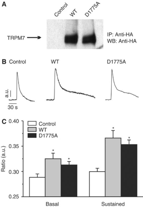
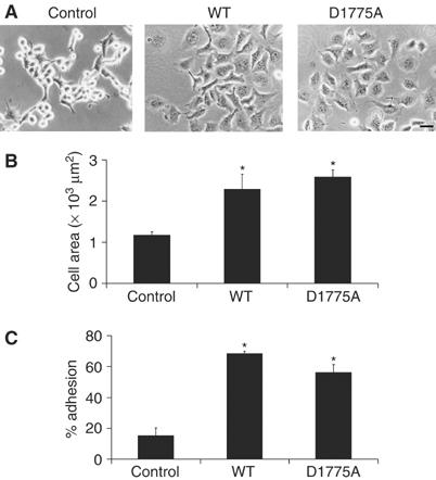
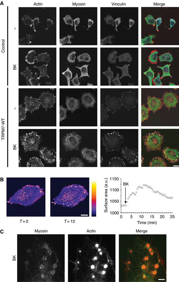
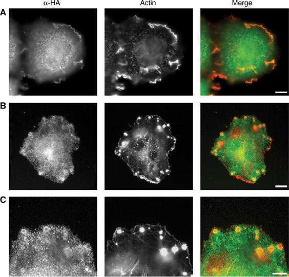
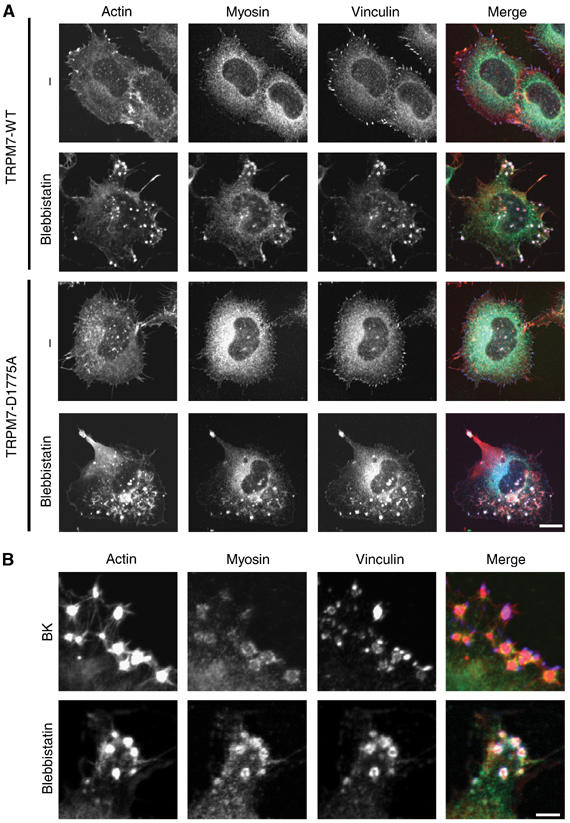
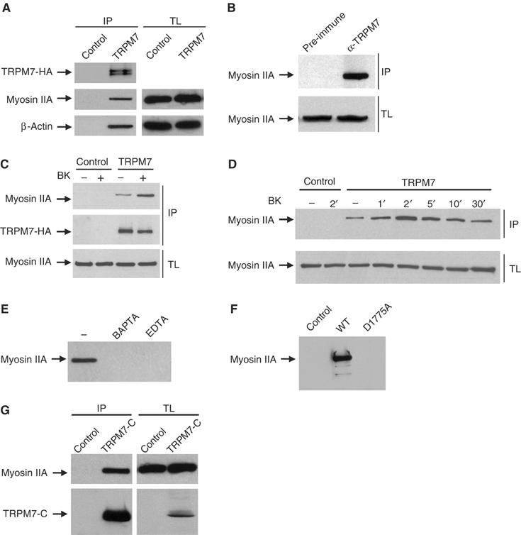
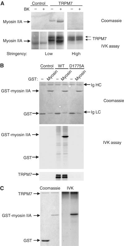
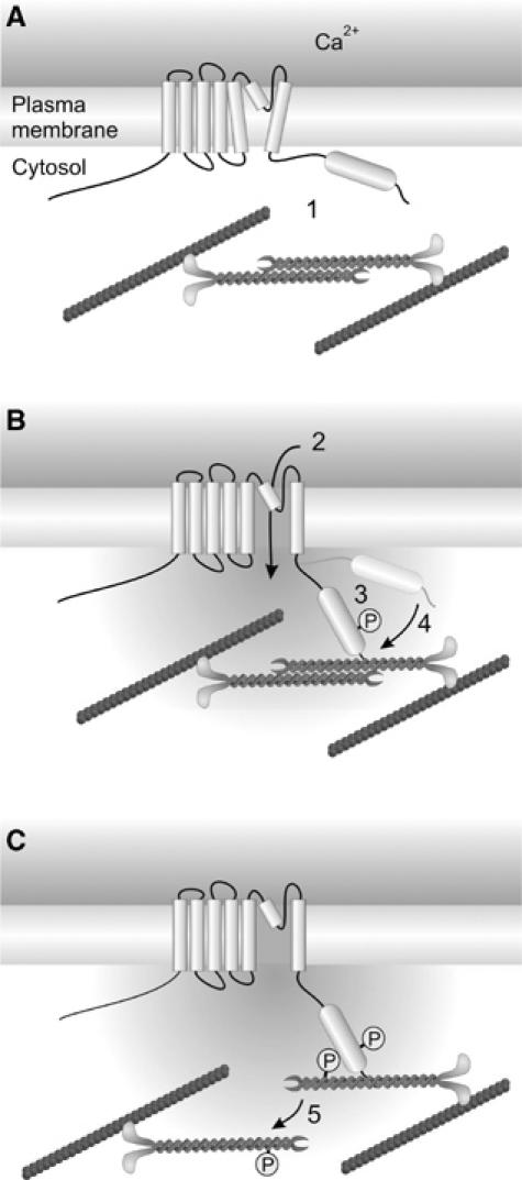
Actomyosin contractility regulates various cell biological processes including cytokinesis, adhesion and migration. While in lower eukaryotes, alpha-kinases control actomyosin relaxation, a similar role for mammalian alpha-kinases has yet to be established. Here, we examined whether TRPM7, a cation channel fused to an alpha-kinase, can affect actomyosin function. We demonstrate that activation of TRPM7 by bradykinin leads to a Ca(2+)- and kinase-dependent interaction with the actomyosin cytoskeleton. Moreover, TRPM7 phosphorylates the myosin IIA heavy chain. Accordingly, low overexpression of TRPM7 increases intracellular Ca2+ levels accompanied by cell spreading, adhesion and the formation of focal adhesions. Activation of TRPM7 induces the transformation of these focal adhesions into podosomes by a kinase-dependent mechanism, an effect that can be mimicked by pharmacological inhibition of myosin II. Collectively, our results demonstrate that regulation of cell adhesion by TRPM7 is the combined effect of kinase-dependent and -independent pathways on actomyosin contractility.


Figures

Figure 1
Figure 1
TRPM7 enhances receptor-operated calcium influx. (A) Expression of TRPM7-WT and TRPM7-D1775A in N1E-115 neuroblastoma cells was detected by IP–Western blot using antibodies against the HA tag. Cells containing the empty vector were used as control in all experiments. (B) TRPM7 expression causes sustained Ca2+ influx following BK stimulation independently of kinase activity. Left panel, typical time course of BK-induced Ca2+ changes in control N1E-115 cells; other panels, BK-induced Ca2+ mobilizations in N1E-115/TRPM7 cells. The sustained influx depends on Ca2+ influx, because it is acutely blocked by extracellular La3+ and Gd3+ (data not shown). (C) Quantification of Ca2+ data for control and TRPM7-overexpressing cells (mean±s.e.m. from at least 20 experiments for each condition). Significant differences (*) are P<0.01 from values obtained in control N1E-115 cells.
Figure 2
Figure 2
TRPM7 overexpression induces cell spreading and increases cell adhesion. (A) Phase-contrast images of control, TRPM7-WT and TRPM7-D1775A-expressing N1E-115 cells. Scale bar=50 μm. (B) Cell surface area covered by N1E-115 cells expressing WT and kinase-dead TRPM7. Cells were stained for F-actin and cell surface area was calculated by quantifying the amount of pixels that exceeded a threshold (n>5). (C) Quantification of cell adhesion of N1E-115 cells expressing WT and kinase-dead TRPM7 (n=3). Significant differences (*) are P<0.01 from values obtained in control N1E-115 cells.
Figure 3
Figure 3
Activation of TRPM7 induces actomyosin reorganization in conjunction with podosome formation. (A) TRPM7 activation induces actomyosin reorganization and formation of podosomes in N1E-115/TRPM7 cells. Cells were either serum starved (0.1% FCS) or stimulated with BK (10 nM, 30 min) and stained for actin (red), myosin IIA heavy chain (green) and vinculin (blue). Cells were analyzed by confocal microscopy. Scale bar=20 μm. (B) Increase in cell surface area and production of podosomes occurs within minutes of BK stimulation in N1E-115/TRPM7 cells. N1E-115/TRPM7 cells expressing GFP-β-actin were followed by confocal microscopy before and after BK stimulation. Cell surface area was measured as described in Materials and methods. Gray values are displayed using the Fire LookUpTable (LUT) of the public domain software Image J (National Institutes of Health, Bethesda, MD, USA). Left panel, a resting cell; middle panel, the same cell 12 min post-BK stimulation; right panel, kinetics of BK-induced cell spreading. A movie is provided as Supplementary data. Scale bar=15 μm. (C) High magnification of myosin lattices found in N1E-115/TRPM7 cells following BK stimulation. N1E-115/TRPM7 cells were transfected with GFP-myosin, stimulated with BK and stained for actin. Scale bar=2 μm.
Figure 4
Figure 4
TRPM7 is present at the cell surface and localizes to cell adhesion structures. N1E-115/TRPM7-WT cells were stimulated with BK (10 nM, 30 min). Subsequently, TRPM7 was detected using anti-HA (3F10) antibody followed by alexa 488-conjugated anti-rat IgG. The actin cytoskeleton was visualized using phalloidin–Texas red. (A) TRPM7 is found in membrane ruffles. Scale bar=10 μm. (B) TRPM7 forms a ring surrounding the actin dense core of podosomes. Scale bar=10 μm. (C) High magnification of TRPM7 rings within podosomes. Scale bar=5 μm. A rat IgG control antibody did not show any specific staining (data not shown).
Figure 5
Figure 5
Regulation of podosome formation by TRPM7 is kinase dependent. N1E-115/TRPM7-WT and N1E-115/TRPM7-D1775A cells were stimulated with BK (10 nM, 30 min) and stained for actin (red), myosin IIA heavy chain (green) and vinculin (blue). Cells were analyzed by confocal microscopy. Scale bar=20 μm.
Figure 6
Figure 6
Inhibition of myosin II function in N1E-115 cells expressing TRPM7-WT and TRPM7-D1775A leads to podosome formation. (A) N1E-115/TRPM7-WT and N1E-115/TRPM7-D1775A cells were incubated in the presence of vehicle control or 50 μM blebbistatin for 30 min. Cells were stained for actin (red), myosin (green) and vinculin (blue) and visualized by confocal microscopy. Scale bar=15 μm. (B) High magnification view shows that the adhesion complexes induced by BK and blebbistatin in N1E-115/TRPM7-WT cells are architecturally similar, as they consist of an actin dense core surrounded by a ring of vinculin and myosin IIA. Scale bar=5 μm.
Figure 7
Figure 7
TRPM7 activation promotes its association via the COOH-terminus with the actomyosin cytoskeleton in a calcium- and kinase-dependent manner. In all experiments (except panel B), N1E-115 control or TRPM7-transduced cells were lysed and incubated with anti-HA-coupled protein G beads for 3 h at 4°C. Proteins in the complex were detected by Western blotting. (A) Co-immunoprecipitation of TRPM7 with β-actin and myosin IIA. Top, detection of TRPM7 with anti-HA antibodies; middle and bottom, detection of myosin IIA and β-actin, respectively, in the immune complexes (IP, left) and total lysates to control for protein levels (TL, right). (B) Endogenous TRPM7 associates with the actomyosin cytoskeleton. PC12 lysates were incubated with preimmune serum or anti-TRPM7 serum and protein complexes were isolated using protein G-Sepharose. The presence of myosin IIA was detected by Western blotting. Top, detection of associated myosin IIA heavy chain in immunoprecipitates; bottom, detection of associated myosin IIA heavy chain in total lysates. (C) Co-immunoprecipitation of TRPM7 with myosin IIA heavy chain pre- and post-BK stimulation (10 nM, 1 min). Top, detection of co-immunoprecipitated myosin IIA; middle, detection of TRPM7 with anti-HA antibodies; bottom, detection of myosin IIA heavy chain in total lysates to control for protein levels. (D) Kinetics of BK-induced TRPM7 association with myosin IIA heavy chain. Cells were stimulated with BK (10 nM) for the indicated times and TRPM7 was immunoprecipitated using anti-HA antibodies. Top, Western blot showing co-immunoprecipitated myosin IIA heavy chain; bottom, detection of myosin IIA heavy chain in total lysates. (E) Interaction between TRPM7 and myosin IIA heavy chain is Ca2+ dependent. N1E-115/TRPM7 cells were preincubated with 10 mM BAPTA or EDTA for 1 min prior to cell lysis and TRPM7 was immunoprecipitated using anti-HA antibodies. (F) TRPM7-D1775A does not interact with myosin IIA heavy chain. TRPM7-WT and -D1775A were immunoprecipitated with anti-HA antibodies and associating myosin IIA heavy chain was detected by Western blotting. (G) The COOH-terminus of TRPM7 interacts with myosin IIA. The soluble COOH-terminus of TRPM7 (aa 1158–1864) was immunoprecipitated from N1E-115/TRPM7-C cells with anti-HA antibodies and associated myosin IIA was detected by Western blotting. Top, detection of associated myosin IIA heavy chain; bottom, detection of TRPM7-C using anti-HA antibodies in immunoprecipitates (IP, left) and total lysates (TL, right).
Figure 8
Figure 8
TRPM7 phosphorylates myosin IIA heavy chain. (A) In vitro kinase assay detecting the phosphorylation of associated myosin IIA heavy chain by TRPM7. TRPM7/myosin IIA heavy chain complexes were isolated before and after stimulation of N1E-115 control and N1E-115/TRPM7 cells with BK (10 nM, 2 min) under low (1% Triton X-100) and high (1% Triton X-100/0.5% deoxycholate/0.1% SDS) stringency conditions. Substrates of TRPM7 were detected by labeling proteins with [γ-32P]ATP, separating products of the in vitro kinase assay by SDS–PAGE (6% gel) followed by autoradiography. Top, Coomassie staining of precipitated proteins; bottom, autoradiogram of phosphorylated proteins. Note that myosin association is lost under high stringency conditions (1% Triton X-100/0.5% deoxycholate/0.1% SDS), whereas TRPM7 autophosphorylation is unaffected. (B) TRPM7 phosphorylates recombinant myosin IIA. TRPM7-WT and -D1775A were immunoaffinity purified from HEK293 cells using anti-HA antibodies under high stringency and mixed with 2 μg of GST or GST-myosin IIA in kinase buffer. To detect phosphorylated myosin IIA, the proteins were separated by SDS–PAGE (12% gel) followed by autoradiography. Top, Coomassie staining of GST-fusion proteins. Note that GST co-migrates with the antibody light chain (Ig LC). Middle, autoradiogram of phosphorylated GST-fusion proteins. Bottom, autoradiogram showing autophosphorylation of WT but not kinase-dead TRPM7. (C) Recombinant TRPM7 kinase phosphorylates myosin IIA. The TRPM7 kinase domain (aa 1403–1864) was produced in E. coli as a fusion with maltose-binding protein and purified on an amylose column. The purified kinase was incubated with 2 μg of GST or GST-myosin IIA in kinase buffer. The proteins were resolved on a 12% SDS–PAGE gel and detected by Coomassie staining. Phosphorylated proteins were visualized by autoradiography. Left, Coomassie staining of gel; right, autoradiogram.
Figure 9
Figure 9
Regulation of actomyosin contractility by TRPM7. (A) In the resting state, TRPM7 is not associated with the actomyosin cytoskeleton (1). (B) Stimulation of cells with PLC-activating agonists induces TRPM7-mediated Ca2+ influx (2) as well as TRPM7 kinase activity. Autophosphorylation (3) promotes a conformational change within TRPM7, allowing for Ca2+-dependent association with myosin IIA (4). (C) Subsequently, TRPM7 phosphorylates the myosin IIA heavy chain, presumably leading to dissociation of myosin filaments and cytoskeletal remodeling (5).

Similar articles

Cited by

References

    1. Burgstaller G, Gimona M (2004) Actin cytoskeleton remodelling via local inhibition of contractility at discrete microdomains. J Cell Sci 117: 223–231 - PubMed
    1. Burridge K, Wennerberg K (2004) Rho and Rac take center stage. Cell 116: 167–179 - PubMed
    1. De la Roche MA, Smith JL, Betapudi V, Egelhoff TT, Cote GP (2002) Signaling pathways regulating Dictyostelium myosin II. J Muscle Res Cell Motil 23: 703–718 - PubMed
    1. DeMali KA, Wennerberg K, Burridge K (2003) Integrin signaling to the actin cytoskeleton. Curr Opin Cell Biol 15: 572–582 - PubMed
    1. Dorovkov MV, Ryazanov AG (2004) Phosphorylation of annexin I by TRPM7 channel-kinase. J Biol Chem 279: 50643–50646 - PubMed

Publication types