. 2005 Aug;1(3):167-73.
doi: 10.1038/nchembio723. Epub 2005 Jul 17.

Small-molecule interaction with a five-guanine-tract G-quadruplex structure from the human MYC promoter

Affiliations

Small-molecule interaction with a five-guanine-tract G-quadruplex structure from the human MYC promoter

Anh Tuân Phan et al. Nat Chem Biol. 2005 Aug.

Erratum in

  • Nat Chem Biol. 2005 Sep;1(4):234

Abstract

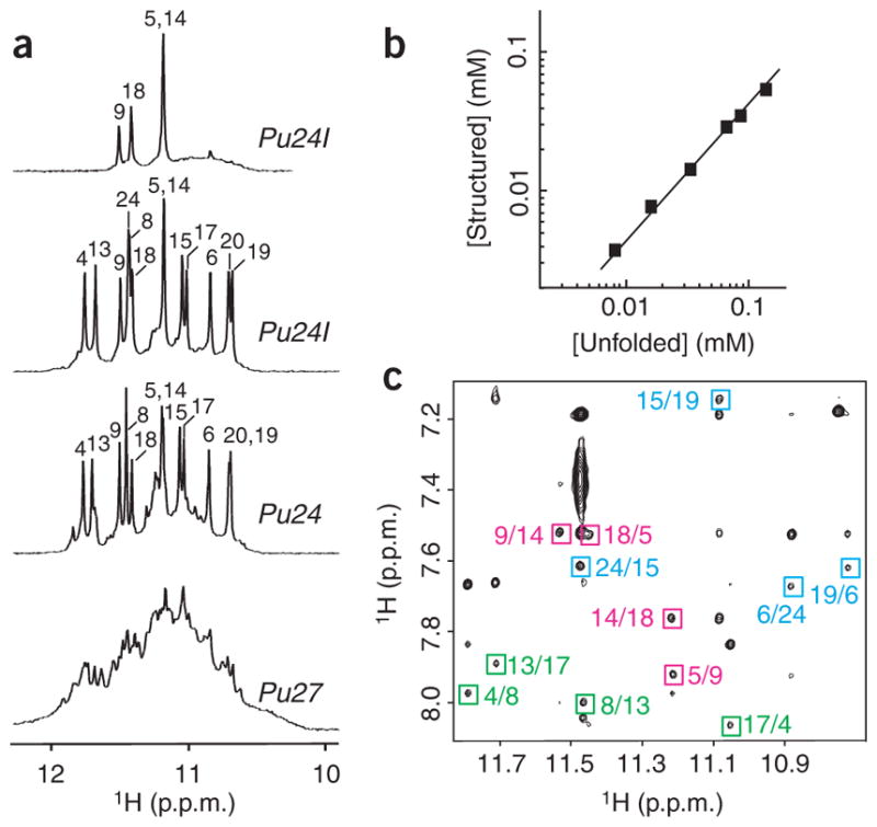
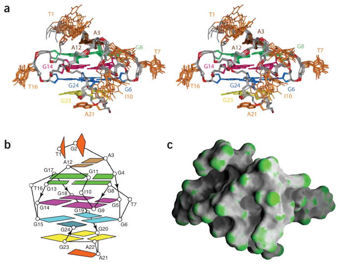
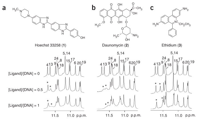
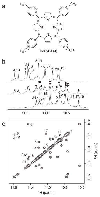
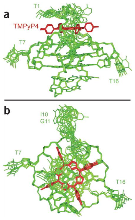
It has been widely accepted that DNA can adopt other biologically relevant structures beside the Watson-Crick double helix. One recent important example is the guanine-quadruplex (G-quadruplex) structure formed by guanine tracts found in the MYC (or c-myc) promoter region, which regulates the transcription of the MYC oncogene. Stabilization of this G-quadruplex by ligands, such as the cationic porphyrin TMPyP4, decreases the transcriptional level of MYC. Here, we report the first structure of a DNA fragment containing five guanine tracts from this region. An unusual G-quadruplex fold, which was derived from NMR restraints using unambiguous model-independent resonance assignment approaches, involves a core of three stacked guanine tetrads formed by four parallel guanine tracts with all anti guanines and a snapback 3'-end syn guanine. We have determined the structure of the complex formed between this G-quadruplex and TMPyP4. This structural information, combined with details of small-molecule interaction, provides a platform for the design of anticancer drugs targeting multi-guanine-tract sequences that are found in the MYC and other oncogenic promoters, as well as in telomeres.


Conflict of interest statement

COMPETING INTERESTS STATEMENT

The authors declare that they have no competing financial interests.

Figures

Figure 1
Figure 1
NMR study of the MYC promoter guanine-rich sequences. (a) The 600 MHz imino proton spectra of Pu27 (bottom), Pu24 (lower middle) and Pu24I in H2O (upper middle) and of Pu24I after 4 h in D2O (top), with unambiguous resonance assignments for both Pu24 and Pu24I listed over the spectra. (b) Determination of stoichiometry of Pu24I by NMR (see Methods). Line of slope 1 is drawn through the data points. (c) NOESY spectrum (mixing time, 200 ms) of Pu24I. Characteristic imino-H8 cross peaks that identify three G-tetrads (colored cyan, magenta and green) are framed and labeled with the number of imino protons in the first position and that of H8 in the second position.
Figure 2
Figure 2
Structure of the Pu24I quadruplex. (a) Stereo view of eight superposed refined structures of the Pu24I quadruplex. The (G6·G24·G15·G19) tetrad is colored cyan, the (G5·G9·G14·G18) tetrad is colored magenta, the (G4·G8·G13·G17) tetrad is colored green, the G20· (A22-G23) triad is colored yellow and the A3·A12 pair is colored brown, with the remaining bases in orange. The backbone is colored white, phosphorus atoms are colored red, and the exocyclic backbone oxygens are omitted in the interest of clarity. (b) Schematic structure of the Pu24I quadruplex. (c) Surface view of a representative refined structure of the Pu24I quadruplex.
Figure 3
Figure 3
Interaction between the Pu24I quadruplex and different ligands as monitored by NMR. (a) Hoechst 33258. (b) Daunomycin. (c) Ethidium. Peaks are labeled with residue numbers. Peaks of G4 and G13 for samples containing ligands are labeled with stars. Pu24I concentration was 0.2 mM.
Figure 4
Figure 4
NMR study of the Pu24I–TMPyP4 complex. (a) The structure of the cationic porphyrin TMPyP4. (b) The interaction between the Pu24I quadruplex and TMPyP4 as monitored by imino proton NMR spectra. Top, middle and bottom spectra are for samples with the [TMPyP4]/[DNA] ratio being 0, 0.5, and 1, respectively. Peaks are labeled with residue numbers. At [TMPyP4]/[DNA] = 0.5, peaks for the Pu24I quadruplex–TMPyP4 complex are marked with black dots. (c) NOESY spectrum (mixing time, 75 ms) of Pu24I in the presence of 50% TMPyP4. Exchange cross-peaks between the free and bound Pu24I quadruplexes are labeled with residue numbers. Pu24I concentration was 0.2 mM for b and 0.4 mM for c.
Figure 5
Figure 5
Six superposed refined structures of the Pu24I quadruplex–TMPyP4 complex. (a) Side view. (b) Top view. Pu24I quadruplex is colored green; TMPyP4 is colored red.

Similar articles

Cited by

References

    1. Marcu KB, Bossone SA, Patel AJ. myc function and regulation. Annu Rev Biochem. 1992;61:809–860. - PubMed
    1. Dang CV. c-Myc target genes involved in cell growth, apoptosis, and metabolism. Mol Cell Biol. 1999;19:1–11. - PMC - PubMed
    1. Pelengaris S, Rudolph B, Littlewood T. Action of Myc in vivo—proliferation and apoptosis. Curr Opin Genet Dev. 2000;10:100–105. - PubMed
    1. Jaattela M. Multiple cell death pathways as regulators of tumour initiation and progression. Oncogene. 2004;23:2746–2756. - PubMed
    1. Slamon DJ, deKernion JB, Verma IM, Cline MJ. Expression of cellular oncogenes in human malignancies. Science. 1984;224:256–262. - PubMed

Publication types

MeSH terms