

Progression of inner ear pathology in Ames waltzer mice and the role of protocadherin 15 in hair cell development
- PMID: 16408167
- PMCID: PMC2504581
- DOI: 10.1007/s10162-005-0024-5
Progression of inner ear pathology in Ames waltzer mice and the role of protocadherin 15 in hair cell development
Abstract
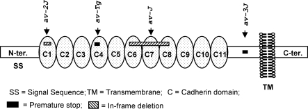
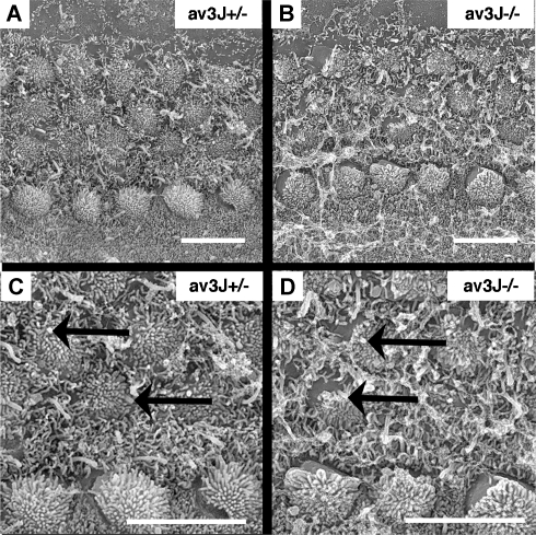
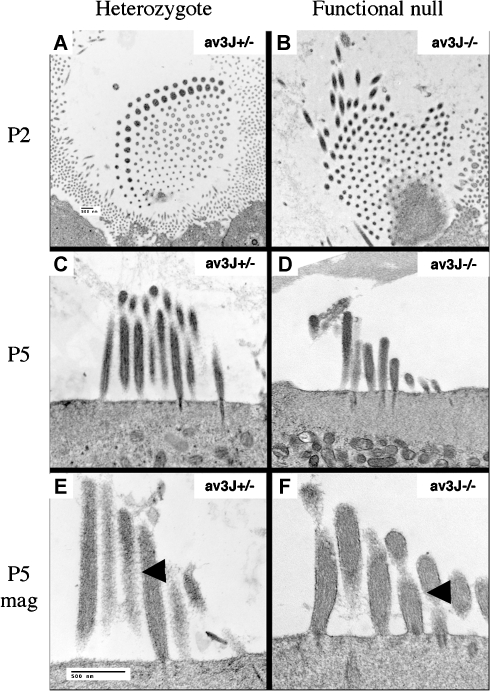
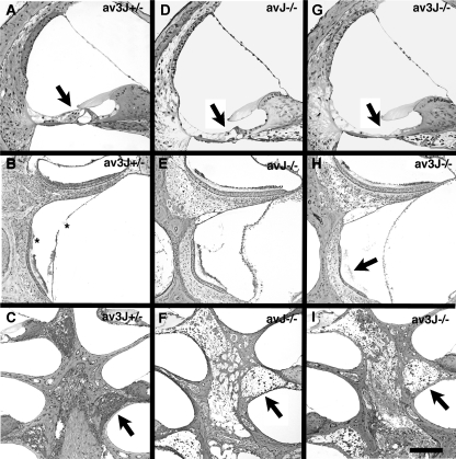
The Ames waltzer (av) mouse mutant exhibits auditory and vestibular abnormalities resulting from mutation of protocadherin 15 (Pcdh15). Ames waltzer has been identified as an animal model for inner ear pathology associated with Usher syndrome type 1F. Studies correlating anatomical phenotype with severity of genetic defect in various av alleles are providing better understanding of the role played by Pcdh15 in inner ear development and of sensorineural abnormalities associated with alterations in Pcdh15 protein structure as a result of gene mutation. In this work we present new findings on inner ear pathology in four alleles of av mice with differing mutations of Pcdh15 as well as varying alterations in inner ear morphology. Two alleles with in-frame deletion mutations (Pcdh15 (av-J) and Pcdh15 (av-2J)) and two presumptive functional null alleles (Pcdh15 (av-3J) and Pcdh15 (av-Tg)) were studied. Light and electron microscopic observations demonstrated that the severity of cochlear and vestibular pathology in these animals correlates positively with the extent of mutation in Pcdh15 from embryonic day 18 (E18) up to 12 months. Electron microscopic analysis of immature ears indicated early abnormalities in the arrangement of stereocilia and the inner and outer hair cell cuticular plates, stereocilia rootlets, and the actin meshwork within the cuticular plate. In severe cases, displacement of the kinocilium and alterations in the shape of the cuticular plate was also observed. Mice harboring in-frame deletion mutations showed less disorganization of stereocilia and cuticular plates in the organ of Corti than the presumptive functional null alleles at P0-P10. A slower progression of pathology was also seen via light microscopy in older animals with in-frame deletions, compared to the presumptive functional null mutations. In summary, our results demonstrate that mutation in Pcdh15 affects the initial formation of stereocilia bundles with associated changes in the actin meshwork within the cuticular plate; these effects are more pronounced in the presumed null mutation compared to mutations that only affect the extracellular domain. The positive correlation of severity of effects with extent of mutation can be seen well into adulthood.
Figures


Similar articles
-
Development of outer hair cells in Ames waltzer mice: mutation in protocadherin 15 affects development of cuticular plate and associated structures.Anat Rec (Hoboken). 2008 Feb;291(2):224-32. doi: 10.1002/ar.20632. Anat Rec (Hoboken). 2008. PMID: 18085631
-
Characterization of a new allele of Ames waltzer generated by ENU mutagenesis.Hear Res. 2005 Apr;202(1-2):161-9. doi: 10.1016/j.heares.2004.09.014. Hear Res. 2005. PMID: 15811708
-
Characterization of vestibular dysfunction in the mouse model for Usher syndrome 1F.J Assoc Res Otolaryngol. 2005 Jun;6(2):106-18. doi: 10.1007/s10162-004-5032-3. Epub 2005 Jun 10. J Assoc Res Otolaryngol. 2005. PMID: 15952048 Free PMC article.
-
Usher I syndrome: unravelling the mechanisms that underlie the cohesion of the growing hair bundle in inner ear sensory cells.J Cell Sci. 2005 Oct 15;118(Pt 20):4593-603. doi: 10.1242/jcs.02636. J Cell Sci. 2005. PMID: 16219682 Review.
-
[Usher syndrome type I and the differentiation of inner ear sensory cells' hair bundles].Med Sci (Paris). 2005 Aug-Sep;21(8-9):737-40. doi: 10.1051/medsci/2005218-9737. Med Sci (Paris). 2005. PMID: 16115459 Review. French.
Cited by
-
Development and regeneration of sensory transduction in auditory hair cells requires functional interaction between cadherin-23 and protocadherin-15.J Neurosci. 2010 Aug 25;30(34):11259-69. doi: 10.1523/JNEUROSCI.1949-10.2010. J Neurosci. 2010. PMID: 20739546 Free PMC article.
-
Mutations in protocadherin 15 and cadherin 23 affect tip links and mechanotransduction in mammalian sensory hair cells.PLoS One. 2011 Apr 21;6(4):e19183. doi: 10.1371/journal.pone.0019183. PLoS One. 2011. PMID: 21532990 Free PMC article.
-
Nerve maintenance and regeneration in the damaged cochlea.Hear Res. 2011 Nov;281(1-2):56-64. doi: 10.1016/j.heares.2011.04.019. Epub 2011 May 10. Hear Res. 2011. PMID: 21596129 Free PMC article. Review.
-
TMPRSS3 expression is limited in spiral ganglion neurons: implication for successful cochlear implantation.J Med Genet. 2022 Dec;59(12):1219-1226. doi: 10.1136/jmg-2022-108654. Epub 2022 Aug 12. J Med Genet. 2022. PMID: 35961784 Free PMC article. Review.
-
A mouse model for nonsyndromic deafness (DFNB12) links hearing loss to defects in tip links of mechanosensory hair cells.Proc Natl Acad Sci U S A. 2009 Mar 31;106(13):5252-7. doi: 10.1073/pnas.0900691106. Epub 2009 Mar 6. Proc Natl Acad Sci U S A. 2009. PMID: 19270079 Free PMC article.
References
-
- {'text': '', 'ref_index': 1, 'ids': [{'type': 'DOI', 'value': '10.1093/hmg/ddg358', 'is_inner': False, 'url': 'https://doi.org/10.1093/hmg/ddg358'}, {'type': 'PubMed', 'value': '14570705', 'is_inner': True, 'url': 'https://pubmed.ncbi.nlm.nih.gov/14570705/'}]}
- Ahmed ZM, Riazuddin S, Ahmad J, Bernstein SL, Guo Y, Sabar MF, Sieving P, Riazuddin S, Griffith AJ, Friedman TB, Belyantseva IA, Wilcox ER. PCDH15 is expressed in the neurosensory epithelium of the eye and ear and mutant alleles are responsible for both USH1F and DFNB23. Hum. Mol. Genet. 12:3215–3223, 2003. - PubMed
-
- {'text': '', 'ref_index': 1, 'ids': [{'type': 'PMC', 'value': 'PMC1460692', 'is_inner': False, 'url': 'https://pmc.ncbi.nlm.nih.gov/articles/PMC1460692/'}, {'type': 'PubMed', 'value': '10430593', 'is_inner': True, 'url': 'https://pubmed.ncbi.nlm.nih.gov/10430593/'}]}
- Alagramam KN, Kwon HY, Cacheiro NLA, Stubbs L, Wright CG, Erway LC, Woychik RP. A new mouse insertional mutation that causes sensorineural deafness and vestibular defects. Genetics 152:1691–1699, 1999. - PMC - PubMed
-
- {'text': '', 'ref_index': 1, 'ids': [{'type': 'DOI', 'value': '10.1016/S0378-5955(00)00152-0', 'is_inner': False, 'url': 'https://doi.org/10.1016/s0378-5955(00)00152-0'}, {'type': 'PubMed', 'value': '10978835', 'is_inner': True, 'url': 'https://pubmed.ncbi.nlm.nih.gov/10978835/'}]}
- Alagramam KN, Zahorsky-Reeves J, Wright CG, Pawlowski KS, Erway LC, Stubbs L, Woychik RP. Neuroepithelial defects of the inner ear in a new allele of the mouse mutation Ames waltzer. Hear. Res. 148:181–191, 2000. - PubMed
-
- {'text': '', 'ref_index': 1, 'ids': [{'type': 'PubMed', 'value': '11138007', 'is_inner': True, 'url': 'https://pubmed.ncbi.nlm.nih.gov/11138007/'}]}
- Alagramam KN, Murcia CL, Kwon HY, Pawlowski KS, Wright CG, Woychik RP. The mouse Ames waltzer hearing-loss mutant is caused by mutation of Pcdh15, a novel protocadherin gene. Nat. Genet. 27:99–102, 2001a. - PubMed
-
- {'text': '', 'ref_index': 1, 'ids': [{'type': 'DOI', 'value': '10.1093/hmg/10.16.1709', 'is_inner': False, 'url': 'https://doi.org/10.1093/hmg/10.16.1709'}, {'type': 'PubMed', 'value': '11487575', 'is_inner': True, 'url': 'https://pubmed.ncbi.nlm.nih.gov/11487575/'}]}
- Alagramam KN, Yuan HJ, Kuehn MH, Murcia CL, Wayne S, Srisailpathy CRS, Lowry RB, Knaus R, Van Laer L, Bernier FP, Schwartz S, Lee C, Morton CC, Mullins RF, Ramesh A, Van Camp G, Hagemen GS, Woychik RP, Smith RJH. Mutations in the novel protocadherin PCDH15 cause Usher syndrome type 1F. Hum. Mol. Genet. 10:1709–1718, 2001b. - PubMed
Publication types
MeSH terms
Substances
Grants and funding
LinkOut - more resources
Full Text Sources
Molecular Biology Databases
Miscellaneous
