

Effect of interleukin-18 on metastasis of mouse osteosarcoma cells
- PMID: 16408211
- PMCID: PMC11030811
- DOI: 10.1007/s00262-005-0097-3
Effect of interleukin-18 on metastasis of mouse osteosarcoma cells
Abstract
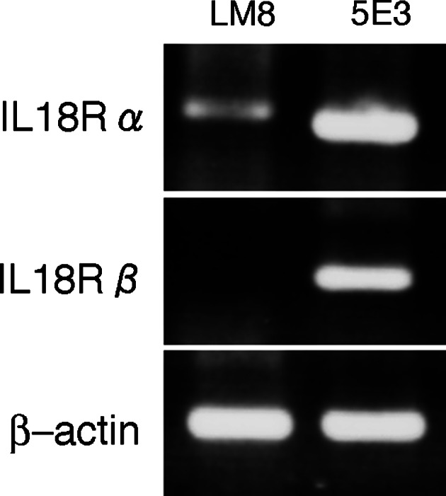
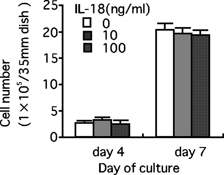
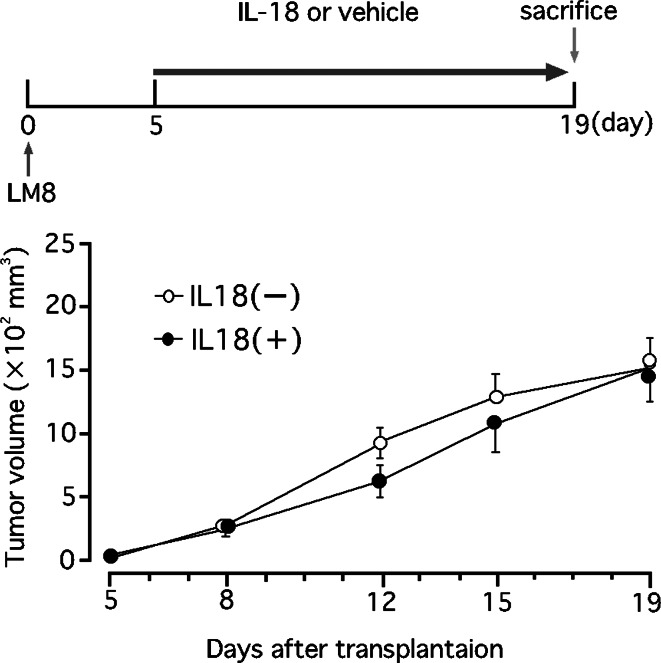
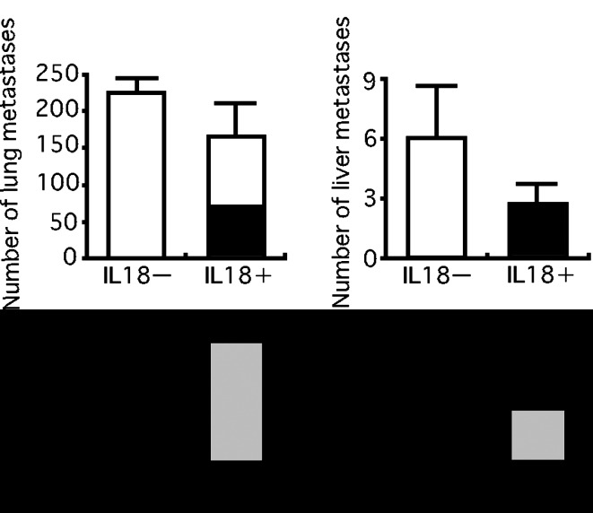
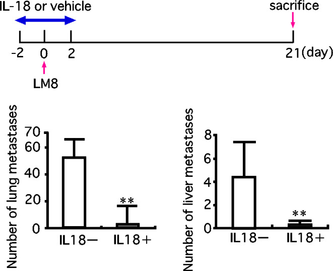
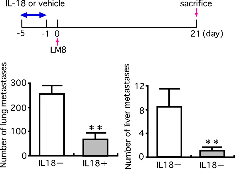
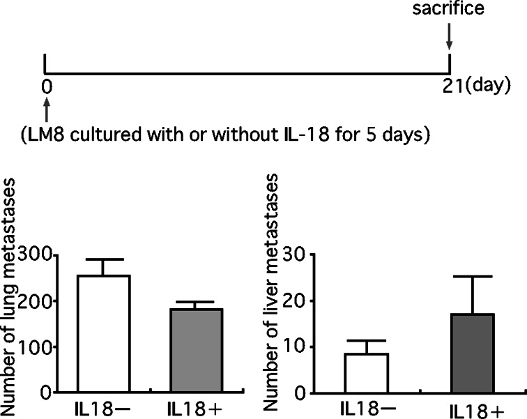
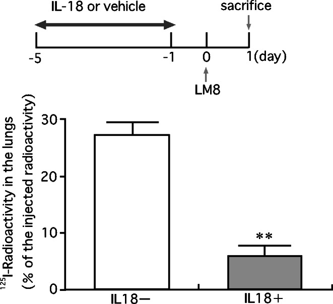
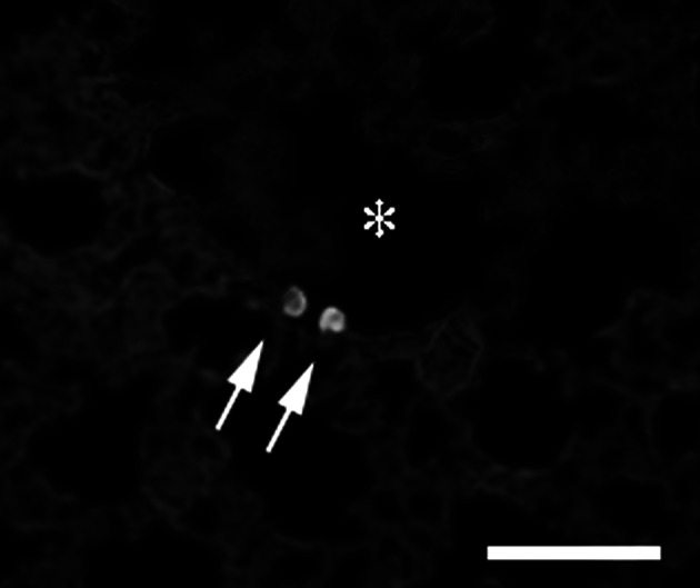
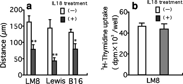
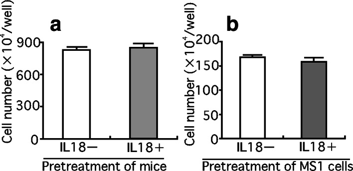
The effect of interleukin-18 (IL-18) on metastasis of highly metastatic LM8 mouse osteosarcoma cells was investigated using nude mice treated with anti-asialo GM1 serum to exclude anti-tumor actions of IL-18 through activation of T and natural killer cells. Injection of LM8 cells which do not express IL-18 receptor beta into a tail vain resulted in the formation of pulmonary and hepatic metastatic foci. Daily injection of mice with IL-18 starting the fifth day from the cell injection had no significant effect on the number of metastatic foci, while five daily injections of IL-18 before and after the cell injection resulted in marked decreases. Culture of LM8 cells with IL-18 for 5 days before the injection into mice produced no significant effect on the number of pulmonary and hepatic metastatic foci. In contrast, pretreatment of mice with IL-18 for 5 days before the cell injection markedly decreased metastatic foci. The retention of LM8 cells in the lung 24 h after their injection was also reduced by the pretreatment of mice with IL-18. Serum obtained from mice pretreated with IL-18 for 5 days suppressed mobility of LM8 cells but IL-18 itself did not. These results suggest that IL-18 inhibits metastasis of LM8 cells partly by inducing a factor(s) in the host which suppresses cell mobility.
Figures
Similar articles
-
Dynamic analysis of lung metastasis by mouse osteosarcoma LM8: VEGF is a candidate for anti-metastasis therapy.Clin Exp Metastasis. 2013 Apr;30(4):369-79. doi: 10.1007/s10585-012-9543-8. Epub 2012 Oct 18. Clin Exp Metastasis. 2013. PMID: 23076771 Free PMC article.
-
Antimetastatic activity of honokiol in osteosarcoma.Cancer. 2012 Apr 15;118(8):2117-27. doi: 10.1002/cncr.26434. Epub 2011 Sep 20. Cancer. 2012. PMID: 21935912
-
Immunotherapy with interleukin-18 in combination with preoperative chemotherapy with ifosfamide effectively inhibits postoperative progression of pulmonary metastases in a mouse osteosarcoma model.Tumour Biol. 2009;30(4):176-84. doi: 10.1159/000236410. Epub 2009 Sep 9. Tumour Biol. 2009. PMID: 19738413
-
Inhibition by interleukin-18 of the growth of Dunn osteosarcoma cells.J Interferon Cytokine Res. 2004 Mar;24(3):161-7. doi: 10.1089/107999004322917007. J Interferon Cytokine Res. 2004. PMID: 15035849
-
Hybrid liposomes inhibit tumor growth and lung metastasis of murine osteosarcoma cells.Cancer Med. 2013 Jun;2(3):267-76. doi: 10.1002/cam4.67. Epub 2013 Mar 22. Cancer Med. 2013. PMID: 23930203 Free PMC article.
Cited by
-
Effect of lentiviral vector-packaged interleukin-18 gene on the malignant behavior of lung cancer.Exp Ther Med. 2020 Jan;19(1):319-326. doi: 10.3892/etm.2019.8204. Epub 2019 Nov 15. Exp Ther Med. 2020. PMID: 31853306 Free PMC article.
-
Gene-Edited Interleukin CAR-T Cells Therapy in the Treatment of Malignancies: Present and Future.Front Immunol. 2021 Jul 27;12:718686. doi: 10.3389/fimmu.2021.718686. eCollection 2021. Front Immunol. 2021. PMID: 34386015 Free PMC article. Review.
-
Molecular alterations as target for therapy in metastatic osteosarcoma: a review of literature.Clin Exp Metastasis. 2011 Jun;28(5):493-503. doi: 10.1007/s10585-011-9384-x. Epub 2011 Apr 2. Clin Exp Metastasis. 2011. PMID: 21461590 Free PMC article. Review.
-
Human IL18-IL2 fusion protein as a potential antitumor reagent by enhancing NK cell cytotoxicity and IFN-γ production.J Cancer Res Clin Oncol. 2012 Oct;138(10):1727-36. doi: 10.1007/s00432-012-1248-5. Epub 2012 Jun 15. J Cancer Res Clin Oncol. 2012. PMID: 22699929 Free PMC article.
-
Relationship of common variants in Interleukin 33 gene with susceptibility and prognosis of osteosarcoma in Han Chinese population.J Cancer. 2019 Jan 29;10(5):1138-1144. doi: 10.7150/jca.29086. eCollection 2019. J Cancer. 2019. PMID: 30854122 Free PMC article.
References
-
- Asai T, Ueda T, Itoh K, Yoshioka K, Aoki Y, Mori S, Yoshikawa H. Establishment and characterization of a murine osteosarcoma cell line (LM8) with high metastatic potential to the lung. Int J Cancer. 1998;76:418–422. doi: 10.1002/(SICI)1097-0215(19980504)76:3<418::AID-IJC21>3.0.CO;2-5. - DOI - PubMed
-
- Cao R, Farnebo J, Kurimoto M, Cao Y. Interleukin-18 acts as angiogenesis and tumor suppressor. FASEB J. 1999;13:2195–2202. - PubMed
-
- Carrascal MT, Mendoza L, Valcarcel M, Salado C, Egilegor E, Telleria N, Vidal-Vanaclocha F, Dinarello CA. Interleukin-18 binding protein reduces B16 melanoma hepatic metastasis by neutralizing adhesiveness and growth factors of sinusoidal endothelium. Cancer Res. 2003;63:491–497. - PubMed
-
- Glasgow LA, Kern ER. Effect of interferon administration on pulmonary osteogenic sarcomas in an experimental murine model. J Natl Cancer Inst. 1981;67:207–212. - PubMed
Publication types
MeSH terms
Substances
LinkOut - more resources
Full Text Sources
Medical
Miscellaneous
