Idiopathic sclerosing orbital pseudotumor in seven cats
- PMID: 16409245
- PMCID: PMC7169322
- DOI: 10.1111/j.1463-5224.2005.00436.x
Idiopathic sclerosing orbital pseudotumor in seven cats
Abstract
Objective: To review the clinical presentation and histopathologic findings on a series of cats with orbital fibrotic disease and compare the data to that of humans with sclerosing orbital pseudotumor.
Animals: A retrospective study was undertaken, which identified tissue samples from seven cats between 1997 and 2002 with a history of orbital mass effect and pathology characterized by fibrous tissue proliferation.
Procedure: Information was obtained from medical records for affected cats, including age, sex, clinical signs, management, and outcome, with histopathology re-examined.
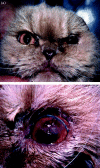
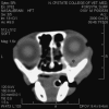
Results: Six of seven cats presented with unilateral orbital involvement that progressed to bilateral orbital disease despite treatment. Onset was insidious, evolving over weeks to months and was associated with fixation of orbital structures. Owners of six of the cats opted for euthanasia because of disease progression and pain. Histopathology of affected orbital tissue included extensive fibrosis with encapsulation of normal tissues without characteristics of neoplasia.
Conclusions: Clinical findings and histopathology of globes and orbital tissues in cats bore many similarities to idiopathic sclerosing orbital pseudotumor in humans. In cats, the prognosis for the globe appears to be poor but an elucidation of the pathogenesis and earlier diagnosis coupled with more aggressive treatment modalities as indicated in humans may be beneficial.
Figures


References
-
- Williams DL, Long RD, Barnett KC. Lacrimal pseudotumour in a young bull terrier. Journal of Small Animal Practice 1998; 39: 30–32. - PubMed
-
- Dziezyc J, Barton CL. Exophthalmia in a cat caused by an eosinophilic infiltrate. Progress in Veterinary and Comparative Ophthalmology 1991; 2: 91–93.
-
- Miller SA, van Der Woerdt A, Bartick TE. Retrobulbar pseudotumor of the orbit in a cat. Journal of the American Veterinary Medical Association 2000; 216: 356–358. - PubMed
-
- Kennerdell JS, Dresner SC. The nonspecific orbital inflammatory syndromes. Survey of Ophthalmology 1984; 29: 93–103. - PubMed
-
- Rootman J, McCarthy M, White V et al. Idiopathic sclerosing inflammation of the orbit: a distinct clinicopathologic entity. Ophthalmology 1994; 101: 570–584. - PubMed
MeSH terms
Substances
LinkOut - more resources
Full Text Sources
Miscellaneous

