. 2006 Jan-Feb;8(1):101-10.
doi: 10.1111/j.1525-142X.2006.05079.x.

Generality of vertebrate developmental patterns: evidence for a dermomyotome in fish

Affiliations

Generality of vertebrate developmental patterns: evidence for a dermomyotome in fish

S H Devoto et al. Evol Dev. 2006 Jan-Feb.

Erratum in

  • Evol Dev. 2006 Mar-Apr;8(2):239

Abstract

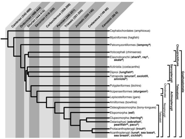
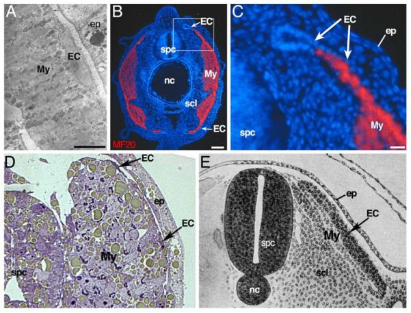
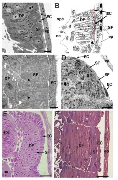
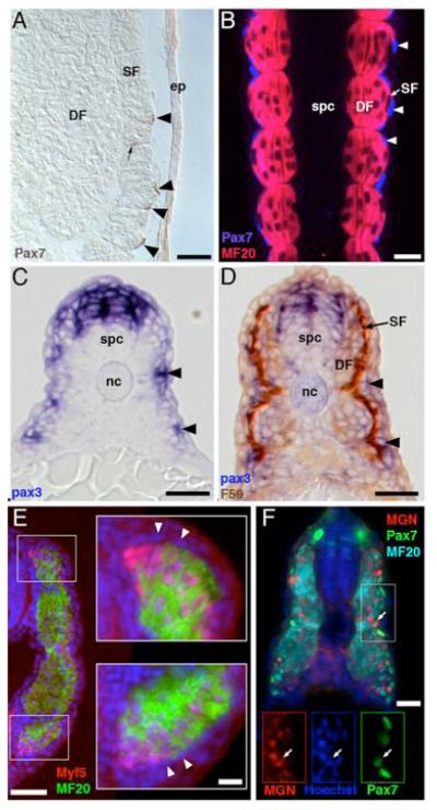
The somitic compartment that gives rise to trunk muscle and dermis in amniotes is an epithelial sheet on the external surface of the somite, and is known as the dermomyotome. However, despite its central role in the development of the trunk and limbs, the evolutionary history of the dermomyotome and its role in nonamniotes is poorly understood. We have tested whether a tissue with the morphological and molecular characteristics of a dermomyotome exists in nonamniotes. We show that representatives of the agnathans and of all major clades of gnathostomes each have a layer of cells on the surface of the somite, external to the embryonic myotome. These external cells do not show any signs of terminal myogenic or dermogenic differentiation. Moreover, in the embryos of bony fishes as diverse as sturgeons (Chondrostei) and zebrafish (Teleostei) this layer of cells expresses the pax3 and pax7 genes that mark myogenic precursors. Some of the pax7-expressing cells also express the differentiation-promoting myogenic regulatory factor Myogenin and appear to enter into the myotome. We therefore suggest that the dermomyotome is an ancient and conserved structure that evolved prior to the last common ancestor of all vertebrates. The identification of a dermomyotome in fish makes it possible to apply the powerful cellular and genetic approaches available in zebrafish to the understanding of this key developmental structure.


Figures

None
1. Phylogenetic relationships among selected groups of extant vertebrates. Cladogram is based on Helfman et al., 1997. with some modifications based on recent analysis of the evolutionary relationships between Petromyzontiformes, Myxiniformes, and the Gnathostomata (Furlong and Holland, 2002; Takezaki et al., 2004). The time of divergence between groups is approximately indicated (Stiassny, et al., 2004). In embryos of all the animals listed in bold there is evidence of a layer of undifferentiated cells external to the embryonic myotome. Selected references for each are as follows: aMaurer, 1894; Nakao, 1977; see Fig. 2A bKaestner, 1892; Maurer, 1906 cKaestner, 1892; Maurer, 1894; Sunier, 1911 dSunier, 1911; see Figs. 2B, C eMaurer, 1906; Sunier, 1911 fMaurer, 1894 gTajbakhsh, 2003; see Fig. 2E hMaurer, 1894; Grimaldi et al., 2004, see Fig. 2D iMaurer, 1894; see Fig. 3A, 4A jSunier, 1911; see Fig. 3B kJohnston, 1993 lWaterman, 1969; see Fig. 4B-D, F mStoiber et al., 1998; Fig. 3C nDal Pai et al., 2000 oMaurer, 1894; Vialleton, 1902; see Figs. 3E, F pSee Fig. 3D qVeggetti et al., 1990 rLopez-Albors et al., 1998; Ramirez-Zarzosa et al., 1995 sSee Fig. 4E
None
2. Representative agnathan, chondrichthyan and sarcopterygian embryos each have a layer of undifferentiated cells external to the myotome. A. External cells in a lamprey, Lampetra japonica. Electron micrograph of a transverse section of a midtrunk myotome of a 5-day lamprey (modified from Nakao, 1977). The myotome is composed of horizontal muscle lamellae, bounded on the lateral surface by undifferentiated external cells which are separated from the epidermis by a large extracellular space containing basal lamina (thick arrow). Scale bar = 5 μm B. External cells in a skate Raja erinacea: transverse section of the anterior tail of an embryo. The myotome is immunolabeled with the MF20 myosin antibody (red), and nuclei with Hoechst 33258 (blue). An apparent epithelial cell layer external to the myotome does not express myosin at this time. Scale bar = 100 μm. C. High magnification view of boxed area in B. The external cells form structures similar to the dorsomedial and ventrolateral lips of the amniote dermomyotome. Scale bar = 25 μm. D. External cells in Xenopus sp. Transverse section of somite 8 in a stage 35 tadpole (modified from Grimaldi et al., 2004). External to the myotome is a thin layer of external cells. E. ’External cells’ in a chick, Gallus gallus: drawing of transverse section through the trunk of an embryo (modified from Lillie, 1919). The chick somite begins as an epithelial sphere and then de-epithelializes as it becomes subdivided into sclerotome, myotome, and an epithelial layer external to the myotome. This epithelial layer was called the dermatome by early investigators, but is now known as the dermomyotome; we have labeled it ‘external cells’ to emphasize its similarity to other vertebrates. EC, external cells; My, myotome; ep, epidermis; scl, sclerotome; spc, spinal cord; nc, notochord.
None
3. Actinopterygian embryos have a layer of undifferentiated cells external to the myotome. All species examined have two layers of differentiated muscle fibers: superficial and deep. A. External cells in the sturgeon Acipenser ruthenus. Semi-thin transverse section posterior to the anal vent of an embryo at hatching (at least 60 somites). External to the superficial fibers is a layer of thin external cells. Scale bar = 2 μm. B. External cells in an eel Muraena sp. Transverse section of 5 days old embryo; drawing modified from (modified from Sunier, 1911). A layer of external cells (pale red) is clearly attached to the myotome and separated from the epidermis. C. External cells in the pearlfish Rutilus frisii meidingeri. Electron micrograph of a segment of the dorsolateral surface of the myotome in a transverse section of a 40 somite stage embryo. External cells form a very thin squamous epithelium and show no evidence of myogenic differentiation. Scale bar = 2 μm. D. External cells in the yellowfin tuna Thunnus albacares. Transverse section of an epaxial quadrant from an embryo at hatching (21 hrs post fertilization). Some flattened ECs are present at the lateral surface of the myotome. Superficial fibers are very small in size and arranged in a discontinuous layer. Scale bar = 5 μm. E. External cells in trout Salmo trutta. Semi-thin transverse section just posterior to the anal vent of an eyed stage trout embryo (55-60 somites). External cells lie over the superficial fibers as in other actinopterygians. However, the cells form an apparent cuboidal epithelium more similar to the external layer in skate (Fig. 1B) and chick (Figure 1C). Scale bar = 25 μm. F. External cells in trout are not elongated and do not show any indications of myogenic differentiation. Semi-thin horizontal section of an eyed stage trout (55-60 somites), just posterior to the anal vent. Superficial and deep fibers have clear myofibril striations. No myofibrils or any other signs of differentiation are found in external cells. Scale bar = 25 μm. EC, external cells; DF, deep fibers; SF, superficial fibers, scl; sclerotome; spc, spinal cord; nc, notochord; ep, epidermis
None
4. Cells external to the embryonic myotome express myogenic pax genes in actinopterygians A. Sturgeon, Acipenser ruthenus, larva, 14 days posthatching. Pax7-positive cells (arrows) external to, and inserted between, the superficial dorsal myotome fibers in near the anal vent, revealed with a Pax7 antibody (brown, DSHB). Note that the stained cells are at positions similar to those of the external cells in Fig. 3A. Scale bar, 25 μm. B. Pax7-positive, myosin-negative cells have the same distribution as external cells in zebrafish (Danio rerio). Zebrafish embryos at the end of segmentation (24h) were double labeled with Pax7 antibody (blue, arrowhead) and a myosin antibody (MF20, red) and optically sectioned in the horizontal plane by confocal microscopy. Pax7-positive nuclei are external to the superficial muscle fibers of the myotome, which are distinguished from the multinucleated deep fibers by being mononucleated. Scale bar, 25μm. C. pax3 is expressed in somitic cells on the external surface of the zebrafish myotome (arrowheads). Embryos at the end of the segmentation period (24h) were labeled for pax3 (Seo et al., 1998) in whole mount by in situ hybridization and then sectioned transversely. pax3 is also expressed in the dorsal neural tube. Scale bar, 25μm. D. pax3-expressing somitic cells (purple, arrowheads) in zebrafish are external to the myotome and do not express the superficial muscle marker slow myosin (F59 antibody, brown). Scale bar, 25μm. E. Myf5-expressing cells are subjacent to the external cell layer in cichlid embryos. Cichlid (Astatotilapia burtoni) embryos at the early pectoral fin bud stage were fixed, and transverse sections from the tail were labeled with antibodies to myosin (MF20, green) and to myf5 (Santa Cruz Biotechnology, green), nuclei were counterstained with Hoechst 33258 (blue). External cells form a continuous layer surrounding the MF20-labeled myotome. Myf5 is expressed in myotomal cells subjacent to the dorsomedial and ventral tips of the external layer. Compare to figure 2B, C, E. Scale bar, 50 μm; inset, 10 μm. F. Pax7 and Myogenin are co-expressed in zebrafish. Zebrafish embryos at the end of segmentation (24h) were sectioned and labeled with antibodies against Myogenin (Santa Cruz Biotechnology, green), Pax7, and myosin (MF20, cyan). Myogenin is expressed in cells in the lateral portion of the myotome, the most lateral of which co-express Myogenin and Pax7. Scale bar, 25 μm. SF, superficial fibers; DF, deep fibers; spc, spinal cord; nc, notochord; ep, epidermis.

Similar articles

Cited by

References

    1. Armand O, Boutineau AM, Mauger A, Pautou MP, Kieny M. Origin of satellite cells in avian skeletal muscles. Arch Anat Microsc Morphol Exp. 1983;72:163–181. - PubMed
    1. Ben-Yair R, Kahane N, Kalcheim C. Coherent development of dermomyotome and dermis from the entire mediolateral extent of the dorsal somite. Development. 2003;130:4325–4336. - PubMed
    1. Ben-Yair R, Kalcheim C. Lineage analysis of the avian dermomyotome sheet reveals the existence of single cells with both dermal and muscle progenitor fates. Development. 2005;132:689–701. - PubMed
    1. Blagden CS, Currie PD, Ingham PW, Hughes SM. Notochord induction of zebrafish slow muscle mediated by Sonic hedgehog. Genes Dev. 1997;11:2163–2175. - PMC - PubMed
    1. Bone Q. Locomotor muscle. In: Hoar WS, Randall DJ, editors. Fish Physiology. Academic Press; New York: 1978. pp. 361–424.

Publication types

MeSH terms

Substances

LinkOut - more resources