. 2006 Jan 12:5:2.
doi: 10.1186/1475-2859-5-2.

Enzymes: An integrated view of structure, dynamics and function

Affiliations

Enzymes: An integrated view of structure, dynamics and function

Pratul K Agarwal. Microb Cell Fact. .

Abstract

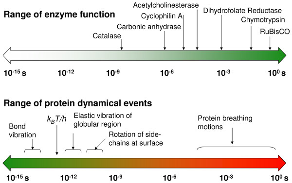
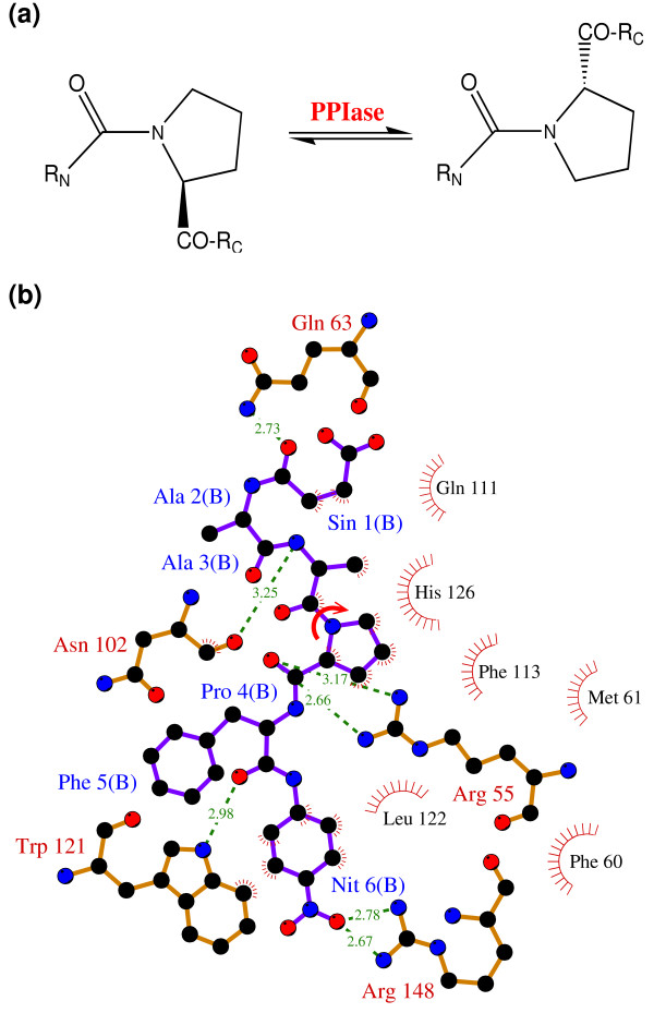
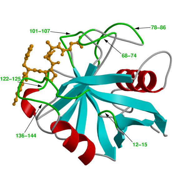
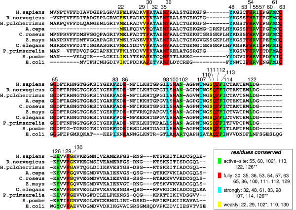
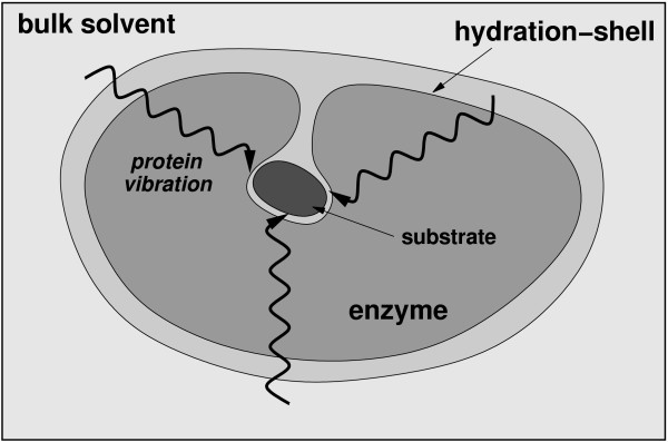
Microbes utilize enzymes to perform a variety of functions. Enzymes are biocatalysts working as highly efficient machines at the molecular level. In the past, enzymes have been viewed as static entities and their function has been explained on the basis of direct structural interactions between the enzyme and the substrate. A variety of experimental and computational techniques, however, continue to reveal that proteins are dynamically active machines, with various parts exhibiting internal motions at a wide range of time-scales. Increasing evidence also indicates that these internal protein motions play a role in promoting protein function such as enzyme catalysis. Moreover, the thermodynamical fluctuations of the solvent, surrounding the protein, have an impact on internal protein motions and, therefore, on enzyme function. In this review, we describe recent biochemical and theoretical investigations of internal protein dynamics linked to enzyme catalysis. In the enzyme cyclophilin A, investigations have lead to the discovery of a network of protein vibrations promoting catalysis. Cyclophilin A catalyzes peptidyl-prolyl cis/trans isomerization in a variety of peptide and protein substrates. Recent studies of cyclophilin A are discussed in detail and other enzymes (dihydrofolate reductase and liver alcohol dehydrogenase) where similar discoveries have been reported are also briefly discussed. The detailed characterization of the discovered networks indicates that protein dynamics plays a role in rate-enhancement achieved by enzymes. An integrated view of enzyme structure, dynamics and function have wide implications in understanding allosteric and co-operative effects, as well as protein engineering of more efficient enzymes and novel drug design.


Figures

Figure 1
Figure 1
The range of time-scales involved in substrate turnover step of enzyme catalyzed reactions and internal protein dynamics are similar. Note the universal frequency factor (kBT/h), which is commonly used in transition state theory; kB is the Boltzmann's constant, T represents the ambient temperature and h is the Planck's constant.
Figure 2
Figure 2
The reaction catalyzed by CypA (a) CypA is a member of a family of enzymes known as PPIase, which catalyze the cis/trans isomerization of peptide bonds N-terminal to proline residues in peptides and proteins (b) The active-site of CypA with a peptide substrate. The shown substrate has the sequence succinyl(Sin)-Ala-Ala-Pro-Phe-p-nitroanilide(Nit) and is labeled as chain B. The red arrow indicates the catalyzed isomerization. Several residues are conserved for their role in enzyme reaction. The dynamical motion of these hydrophobic and hydrophilic residues is linked to the substrate turnover step [10–12]. The green lines indicate hydrogen bonds between substrate and enzyme, while the hydrophobic interactions are depicted by small red radiating lines.
Figure 3
Figure 3
Three-dimensional structure of CypA. Protein secondary structure is represented with cyan arrows (β-sheets) and red helices (α-helices) based on crystal structure from Zhao and Ke (PDB code: 1RMH) [29]. The green labeled regions are flexible surface loops, showing large displacements in X-ray structures (large temperature factors) and NMR relaxation studies. A peptide substrate is shown as orange ball-and-stick model.
Figure 4
Figure 4
Genomic analysis for sequence conservation of CypA. Full analysis was performed on 50 sequences from species ranging from bacteria to human, results from 10 representative sequences are listed above. 17 of 165 amino acid residues in the human CypA sequence were found to be conserved in all 50 PPIases sequences examined: Pro30; Asn35; Phe36; Phe53; His54; Arg55; Ile57; Phe60; Gln63; Gly65; Glu86; Met100; Gln111; Phe112; Phe113; Leu122; and Phe129. Eight additional residues were found to be strongly conserved: Thr32; Tyr48; Met61; Phe83; Leu98; Thr107; Ile114 and His126. Five other residues were found to be weakly conserved: Phe22; Val29; Asn102; Ser110 and Gly130. Conserved active-site residues (fully conserved Arg55, Phe60, Leu122; strongly conserved His126**; and weakly conserved Asn102*) are shown with green background. Red background shows fully conserved residues distal to the active-site; residues with cyan background are strongly conserved; and residues with yellow background are weakly conserved.
Figure 5
Figure 5
Schematic illustration of free energy profile for an enzymatic reaction. Protein dynamics can influence reaction rates in two possible ways; by altering height of the activation free energy barrier (ΔG) and transmission coefficient (κ). kB is the Boltzmann's constant, T is the temperature, h is the Planck's constant and kTST represents the transition state theory reaction rate.
Figure 6
Figure 6
A network of coupled protein vibration promoting catalysis in cyclophilin A [10]. Alternate pathways by which protein dynamics impacts the enzyme reaction are depicted based on 3 protein vibrational modes coupled to the reaction. Loops colored in red and residues indicated by ball-and-stick show largest displacements in vibrational modes coupled to the substrate turnover step. The yellow arrows represent the network pathway from outside of the enzyme to the active-site. Reprinted with permission from Agarwal et al., Biochemistry (2004) 43, 10605–10618. Copyright 2004 American Chemical Society.
Figure 7
Figure 7
Effect of additional kinetic energy in selective protein vibrational modes (a) Increased amount of kinetic energy in a mode coupled to the enzyme reaction allows the trajectory to cross the barrier successfully from the reactant side to the product side. The solid black curve represents the native trajectory (no additional kinetic energy), and δ indicates the fraction of system kinetic energy added to the protein vibrational mode. (b)-(d) representative trajectories from simulations with increased kinetic energy in network protein vibrational modes and a non promoting mode. 2% of system kinetic energy was added to protein vibrations mode. Five representative trajectories from each mode are shown in different colors. Not all protein vibrational modes show increased barrier recrossing; much less effect on the barrier crossing is seen in a mode not coupled to the reaction [12]. Reprinted with permission from Agarwal et al., J. Am. Chem. Soc. (2005) 127, 15248–15256. Copyright (2005) American Chemical Society.
Figure 8
Figure 8
Effect of additional kinetic energy in first solvation shell of an enzyme. (a) Kinetic energy is transferred from the solvent to the protein residues, as indicated by increasing energy in the protein regions (up to 5 Å from protein surface, and between 5 Å and 8 Å from the surface) (b) Two otherwise non-productive regular trajectories (solid lines) become productive (broken lines) due to transfer of energy from the solvent to residues forming parts of the protein vibrations network. The corresponding trajectories are indicated by squares and circles [12]. Reprinted with permission from Agarwal et al., J. Am. Chem. Soc. (2005) 127, 15248–15256. Copyright (2005) American Chemical Society.
Figure 9
Figure 9
A schematic representation of the integrated view of enzyme structure, dynamics and function. Enzyme structure and internal protein dynamics events play a role in the catalytic step. Conserved residues from the surface to the active-site participate in network of protein motions or vibrations that promotes catalysis. The surface residues are coupled to the thermo-dynamical fluctuations of the solvent, and possibly play a role in transfer of energy from solvent to the protein.

Similar articles

Cited by

References

    1. Neet KE. Enzyme catalytic power minireview series. J Biol Chem. 1998;273:25527–25528. doi: 10.1074/jbc.273.40.25527. - DOI - PubMed
    1. Radzicka A, Wolfenden R. A Proficient Enzyme. Science. 1995;267:90–93. - PubMed
    1. Kraut J. How Do Enzymes Work. Science. 1988;242:533–540. - PubMed
    1. Knowles JR. Enzyme Catalysis - Not Different, Just Better. Nature. 1991;350:121–124. doi: 10.1038/350121a0. - DOI - PubMed
    1. Fischer E. Ber Dtsch Chem Ges. 1894. p. 3189.