. 2006 Jan 12:3:4.
doi: 10.1186/1742-4690-3-4.

Intracytoplasmic maturation of the human immunodeficiency virus type 1 reverse transcription complexes determines their capacity to integrate into chromatin

Affiliations

Intracytoplasmic maturation of the human immunodeficiency virus type 1 reverse transcription complexes determines their capacity to integrate into chromatin

Sergey Iordanskiy et al. Retrovirology. .

Abstract

Background: The early events of the HIV-1 life cycle include entry of the viral core into target cell, assembly of the reverse transcription complex (RTCs) performing reverse transcription, its transformation into integration-competent complexes called pre-integration complexes (PICs), trafficking of complexes into the nucleus, and finally integration of the viral DNA into chromatin. Molecular details and temporal organization of these processes remain among the least investigated and most controversial problems in the biology of HIV.

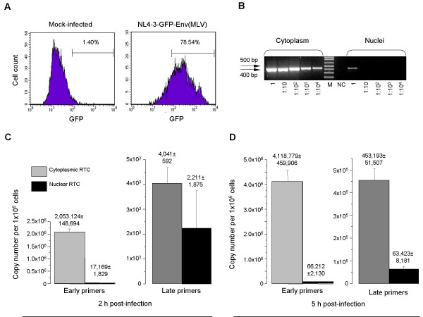
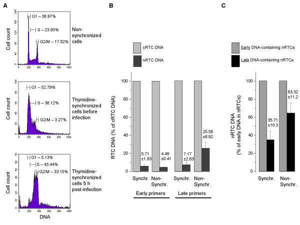
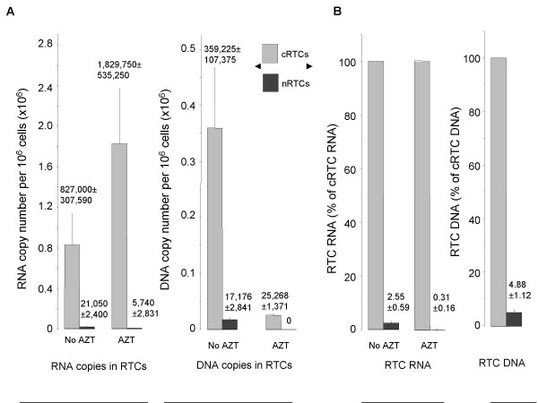
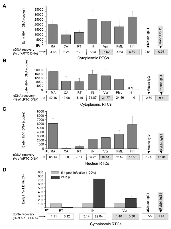
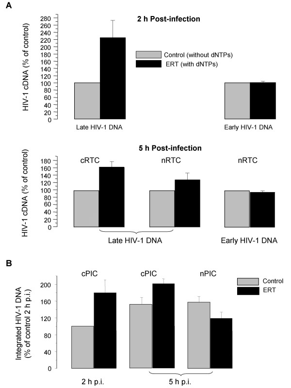
Results: To quantitatively evaluate maturation and nuclear translocation of the HIV-1 RTCs, nucleoprotein complexes isolated from the nucleus (nRTC) and cytoplasm (cRTC) of HeLa cells infected with MLV Env-pseudotyped HIV-1 were analyzed by real-time PCR. While most complexes completed reverse transcription in the cytoplasm, some got into the nucleus before completing DNA synthesis. The HIV-specific RNA complexes could get into the nucleus when reverse transcription was blocked by reverse transcriptase inhibitor, although nuclear import of RNA complexes was less efficient than of DNA-containing RTCs. Analysis of the RTC nuclear import in synchronized cells infected in the G2/M phase of the cell cycle showed enrichment in the nuclei of RTCs containing incomplete HIV-1 DNA compared to non-synchronized cells, where RTCs with complete reverse transcripts prevailed. Immunoprecipitation assays identified viral proteins IN, Vpr, MA, and cellular Ini1 and PML associated with both cRTCs and nRTCs, whereas CA was detected only in cRTCs and RT was diminished in nRTCs. Cytoplasmic maturation of the complexes was associated with increased immunoreactivity with anti-Vpr and anti-IN antibodies, and decreased reactivity with antibodies to RT. Both cRTCs and nRTCs carried out endogenous reverse transcription reaction in vitro. In contrast to cRTCs, in vitro completion of reverse transcription in nRTCs did not increase their integration into chromatin.

Conclusion: These results suggest that RTC maturation occurs predominantly in the cytoplasm. Immature RTCs containing RT and incomplete DNA can translocate into the nucleus during mitosis and complete reverse transcription, but are defective for integration.


Figures

Figure 1
Figure 1
Analysis of nucleo-cytoplasmic distribution of HIV-1 RTCs.HeLa cells were spinoculated with MLV Env-pseudotyped NL4-3 or NL4-3-GFP HIV-1. A. HeLa cells infected with GFP-expressing HIV-1 were analyzed by FACS 48 h after infection. Percentage of GFP-positive cells was counted using CellQuest software. B. PCR analysis of the purity of nuclear extracts. Cytoplasmic and nuclear extracts were prepared from the same number of cells (1 × 10⁶) and total DNA was isolated. Undiluted and diluted (1:10, 1:102, 1:103, and 1:104) DNA samples were analyzed by PCR using primers specific for mitochondrial DNA. M – DNA molecular size marker, NC – negative control (H2O). C,D. Real-time PCR analysis of nuclear and cytoplasmic RTCs. DNA isolated from cytoplasmic and nuclear RTCs 2 h (C) and 5 h (D) after spinoculation was analyzed in triplicate with primers specific for early or late HIV-1 DNA using SYBR Green qPCR. Serial dilutions of DNA from 8E5 cells were used as quantitative standards. Results are presented as mean ± SD.
Figure 2
Figure 2
Quantitative analysis of nuclear translocation of HIV-1 RTCs in synchronized cells. A. Cell cycle distribution of control, non-synchronized HeLa cells (upper panel), and cells pre-treated with 2 mM thymidine was measured by flow cytometric analysis before spinoculation (middle panel) and 5 h after spinoculation (lower panel). Percentage of cells at different phases of the cell cycle was counted using CellQuest software. B,C. Nuclear translocation of HIV-1 RTCs. HIV-1 DNA was purified from cytoplasmic and nuclear HIV-1 complexes 5 h after infection of synchronized and non-synchronized HeLa cells. Triplicate samples were analyzed by real-time PCR with primers specific for early and late HIV-1 DNA by measuring SYBR Green fluorescence. Values are means ± SD. Panel B shows percentage of nRTC DNA relative to DNA from cRTCs. Panel C represents percentage of late DNA from nRTCs relative to early nRTC DNA.
Figure 3
Figure 3
Nuclear translocation of RNA and DNA containing HIV-1 PICs. DNA and RNA were purified from cytoplasmic and nuclear HIV-1 complexes 5 h after infection of HeLa cells in the presence or absence of AZT (3 μM). Triplicate samples were analyzed by real-time PCR with primers specific for late HIV-1 DNA by measuring SYBR Green fluorescence. Results are presented as mean ± SD. A. Absolute values of nuclear and cytoplasmic HIV-1 DNA and RNA in RTCs. B. Percentage of nuclear RNA or DNA relative to cytoplasmic RNA or DNA, respectively.
Figure 4
Figure 4
Analysis of protein composition of cytoplasmic and nuclear RTCs. cRTCs and nRTCs purified 5 h after infection were immunoprecipitated using the indicated antibodies and Protein G Sepharose. DNA was isolated from immune complexes and analyzed by real-time PCR as in Fig. 1. DNA recovered in immunoprecipitated RTCs as percentage of total HIV-1 DNA detected in the cRTCs is indicated under the histogram columns. DNA recovery for isotype control antibodies is shown on the right. DNA recovery for mouse mAb is shown in open boxes, for rabbit polyclonal antibodies – in shaded boxes. A,B. Immunoprecipitated cRTCs were analyzed using primers specific for early (A) and late (B) reverse transcription products. N.d. – not done. Results are mean ± SD of triplicate determinations, except for late DNA analysis of anti-MA-precipitated complexes, which was done only once. One representative experiment out of 4 performed is shown. C. Experiment was performed as in A, except that nRTCs were analyzed. Low sensitivity of primers specific for late HIV-1 DNA precluded their use for analysis of nRTCs. Results are mean ± SD of triplicate determinations. One representative experiment out of 4 performed is shown. D. Temporal analysis of cRTCs. Results are mean ± SD of triplicate determinations. One representative experiment out of 3 performed is shown.
Figure 5
Figure 5
Quantitative PCR analysis of ERT activity and integration of cytoplasmic and nuclear RTCs. A. ERT activity of cRTCs and nRTCs isolated 2 h and 5 h post-infection. cRTCs and nRTCs were normalized according to strong-stop (early) HIV-1 DNA content measured by real-time PCR. ERT reaction was performed in duplicate as described in the text. HIV-1 DNA was quantified by real-time PCR. HIV-1 DNA in RTCs incubated without dNTPs (control) was taken as 100%. Results are presented as mean ± SE. B. Quantitative PCR analysis of PIC integration into chromatin. cPICs and nPICs after the ERT reaction performed with or without (control) dNTPs were incubated in triplicate with chromatin samples. DNA was purified and analyzed by Alu-LTR-based real-time nested PCR [29]. Integration efficiency was evaluated relative to integration of cPIC isolated 2 h p.i. Results are presented as mean ± SD.

Similar articles

Cited by

References

    1. Farnet CM, Haseltine WA. Integration of human immunodeficiency virus type 1 DNA in vitro. Proc Natl Acad Sci U S A. 1990;87:4164–4168. - PMC - PubMed
    1. Nisole S, Saib A. Early steps of retrovirus replicative cycle. Retrovirology. 2004;1:9. doi: 10.1186/1742-4690-1-9. - DOI - PMC - PubMed
    1. Fassati A, Goff SP. Characterization of intracellular reverse transcription complexes of human immunodeficiency virus type 1. J Virol. 2001;75:3626–3635. doi: 10.1128/JVI.75.8.3626-3635.2001. - DOI - PMC - PubMed
    1. Bukrinsky MI, Sharova N, McDonald TL, Pushkarskaya T, Tarpley WG, Stevenson M. Association of integrase, matrix, and reverse transcriptase antigens of human immunodeficiency virus type 1 with viral nucleic acids following acute infection. Proc Natl Acad Sci U S A. 1993;90:6125–6129. - PMC - PubMed
    1. Heinzinger NK, Bukrinsky MI, Haggerty SA, Ragland AM, Kewalramani V, Lee MA, Gendelman HE, Ratner L, Stevenson M, Emerman M. The Vpr protein of human immunodeficiency virus type 1 influences nuclear localization of viral nucleic acids in nondividing host cells. Proc Natl Acad Sci U S A. 1994;91:7311–7315. - PMC - PubMed

Publication types