. 2006 Jan 12:6:4.
doi: 10.1186/1472-6750-6-4.

Development of a new bicistronic retroviral vector with strong IRES activity

Affiliations

Development of a new bicistronic retroviral vector with strong IRES activity

Patrick Martin et al. BMC Biotechnol. .

Abstract

Background: Internal Ribosome Entry Site (IRES)-based bicistronic vectors are important tools in today's cell biology. Among applications, the expression of two proteins under the control of a unique promoter permits the monitoring of expression of a protein whose biological function is being investigated through the observation of an easily detectable tracer, such as Green Fluorescent Protein (GFP). However, analysis of published results making use of bicistronic vectors indicates that the efficiency of the IRES-controlled expression can vary widely from one vector to another, despite their apparent identical IRES sequences. We investigated the molecular basis for these discrepancies.

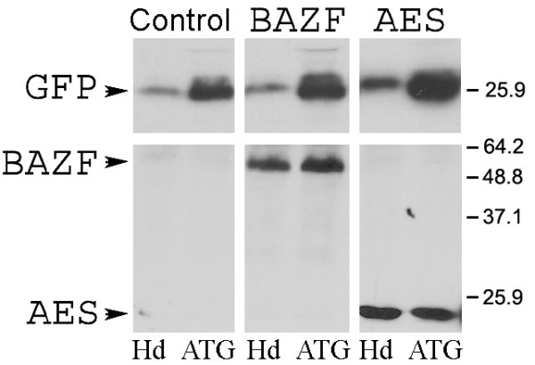
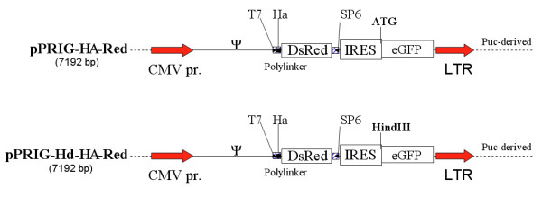
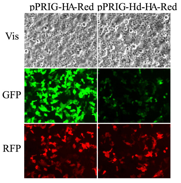
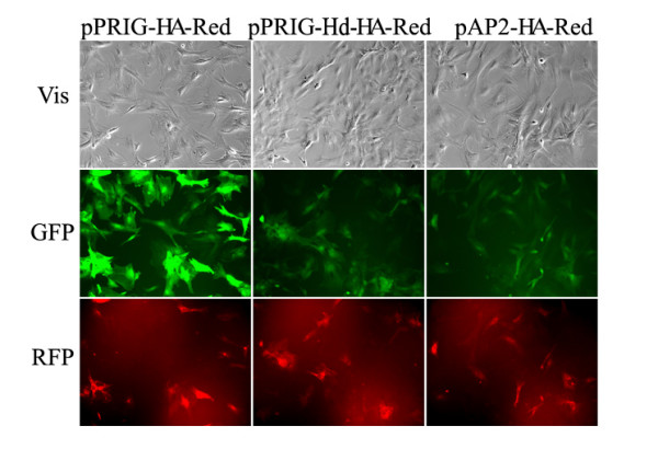
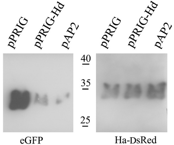
Results: We observed up to a 10 fold difference in IRES-controlled expression from distinct bicistronic expression vectors harboring the same apparent IRES sequences. We show that the insertion of a HindIII site, in place of the initiating AUG codon of the wild type EMCV IRES, is responsible for the dramatic loss of expression from the second cistron, whereas expression from the first cistron remains unaffected. Thus, while the replacement of the authentic viral initiating AUG by a HindIII site results in the theoretical usage of the initiation codon of the HindIII-subcloned cDNA, the subsequent drop of expression dramatically diminishes the interest of the bicistronic structure. Indeed, insertion of the HindIII site has such a negative effect on IRES function that detection of the IRES-controlled product can be difficult, and sometimes even below the levels of detection. It is striking to observe that this deleterious modification is widely found in available IRES-containing vectors, including commercial ones, despite early reports in the literature stating the importance of the integrity of the initiation codon for optimal IRES function.

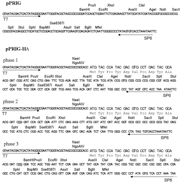
Conclusion: From these observations, we engineered a new vector family, pPRIG, which respects the EMCV IRES structure, and permits easy cloning, tagging, sequencing, and expression of any cDNA in the first cistron, while keeping a high level of expression from its IRES-dependent second cistron (here encoding eGFP).


Figures

Figure 1
Figure 1
Western blot analysis of HEK 293T cell lysates. Cells were transfected with pMigR-Hd and pMigR-ATG (Control), or pMigR-Hd-BAZF and pMigR-ATG -BAZF (BAZF), or pMigR-Hd-AES and pMigR-ATG-AES (AES). Hd: pMigR-Hd based vectors, ATG: pMigR-ATG based vectors. GFP, BAZF and AES: translation products of the corresponding cDNAs. Molecular weight markers are indicated.
Figure 2
Figure 2
Structure of the four pPRIG vector polylinkers. pPRIG is the prototype polylinker, while the three pPRIG-HA vectors contain additional 1-nucleotide frame-shifted coding sequences for HA-tagging 5' of the polylinker sequence. Any insert can thus be cloned either in-frame downstream of this HA-tag, or directly into the pPRIG, without the HA-tag.
Figure 3
Figure 3
Schematic representation of the pPRIG-HA-Red and pPRIG-Hd-HA-Red retroviral vectors. Top drawing illustrates pPRIG-HA-Red, in which the coding sequence of DsRed has been cloned in the polylinker, while pPRIG-Hd-HA-Red is the same exact construct except that the IRES initiation codon is modified into a HindIII site (see Table 1). The psi letter (Ψ) is the MLV encapsidation sequence, the CMV promoter drives the expression of the bicistronic RNA, the hatched boxes flanking DsRed correspond to the polylinker, encompassing the T7 and SP6 sequences and the HA coding sequence, the IRES corresponds to the EMCV-derived IRES fragment, eGFP is the coding sequence for green fluorescent protein used to track the first cistron expression, and the LTR corresponds to the MLV long terminal repeat. The hatched line symbolizes Puc-derived plasmid backbone.
Figure 4
Figure 4
Comparison of transient pPRIG-HA-Red and pPRIGw-HA-Red expressions. HEK 293T cells were transfected with each vector, and the cells were photographed 48 h later. Vis: phase contrast image. Exposure time is the same for the two GFP images, and for the two RFP images. Fluorescence signals are thus a direct reflection of protein expression levels.
Figure 5
Figure 5
Comparison of stable pPRIG-HA-Red, pPRIG-Hd-HA-Red and pAP2-HA-Red expressions. HEK 293T cells were cotransfected with each vector and helper constructs. Viral supernatants were collected after 2 days and applied to primary rat embryo fibroblasts. The REF cells were photographed two weeks post-transduction. Vis: phase contrast image. Exposure time was the same for the three GFP images, as well as for the three RFP images. Fluorescence intensities are thus a direct reflection of protein expression levels.
Figure 6
Figure 6
Comparison of the pPRIG-HA-Red (pPRIG), pPRIG-Hd-HA-Red (pPRIG-Hd) and pAP2-HA-Red (pAP2) GFP and RFP protein expression by Western blot analysis. Equivalent amounts of cell lysates containing the eGFP and HA-DsRed expressed from the indicated vectors were loaded in duplicate, ran on SDS-PAGE, and analyzed with an anti GFP serum (left panel) and an anti-HA serum (right panel).

References

    1. Chambard JC, Pognonec P. A reliable way of obtaining stable inducible clones. Nucleic Acids Res. 1998;26:3443–3444. doi: 10.1093/nar/26.14.3443. - DOI - PMC - PubMed
    1. Ngoi SM, Chien AC, Lee CG. Exploiting internal ribosome entry sites in gene therapy vector design. Curr Gene Ther. 2004;4:15–31. doi: 10.2174/1566523044578095. - DOI - PubMed
    1. Jang SK, Wimmer E. Cap-independent translation of encephalomyocarditis virus RNA: structural elements of the internal ribosomal entry site and involvement of a cellular 57-kD RNA-binding protein. Genes Dev. 1990;4:1560–1572. - PubMed
    1. Ohlmann T, Jackson RJ. The properties of chimeric picornavirus IRESes show that discrimination between internal translation initiation sites is influenced by the identity of the IRES and not just the context of the AUG codon. Rna. 1999;5:764–778. doi: 10.1017/S1355838299982158. - DOI - PMC - PubMed
    1. Galipeau J, Li H, Paquin A, Sicilia F, Karpati G, Nalbantoglu J. Vesicular stomatitis virus G pseudotyped retrovector mediates effective in vivo suicide gene delivery in experimental brain cancer. Cancer Res. 1999;59:2384–2394. - PubMed

Publication types

Substances

LinkOut - more resources