. 2006 Jan 13:3:5.
doi: 10.1186/1742-4690-3-5.

SUV39H1 interacts with HTLV-1 Tax and abrogates Tax transactivation of HTLV-1 LTR

Affiliations

SUV39H1 interacts with HTLV-1 Tax and abrogates Tax transactivation of HTLV-1 LTR

Koju Kamoi et al. Retrovirology. .

Abstract

Background: Tax is the oncoprotein of HTLV-1 which deregulates signal transduction pathways, transcription of genes and cell cycle regulation of host cells. Transacting function of Tax is mainly mediated by its protein-protein interactions with host cellular factors. As to Tax-mediated regulation of gene expression of HTLV-1 and cellular genes, Tax was shown to regulate histone acetylation through its physical interaction with histone acetylases and deacetylases. However, functional interaction of Tax with histone methyltransferases (HMTase) has not been studied. Here we examined the ability of Tax to interact with a histone methyltransferase SUV39H1 that methylates histone H3 lysine 9 (H3K9) and represses transcription of genes, and studied the functional effects of the interaction on HTLV-1 gene expression.

Results: Tax was shown to interact with SUV39H1 in vitro, and the interaction is largely dependent on the C-terminal half of SUV39H1 containing the SET domain. Tax does not affect the methyltransferase activity of SUV39H1 but tethers SUV39H1 to a Tax containing complex in the nuclei. In reporter gene assays, co-expression of SUV39H1 represses Tax transactivation of HTLV-1 LTR promoter activity, which was dependent on the methyltransferase activity of SUV39H1. Furthermore, SUV39H1 expression is induced along with Tax in JPX9 cells. Chromatin immunoprecipitation (ChIP) analysis shows localization of SUV39H1 on the LTR after Tax induction, but not in the absence of Tax induction, in JPX9 transformants retaining HTLV-1-Luc plasmid. Immunoblotting shows higher levels of SUV39H1 expression in HTLV-1 transformed and latently infected cell lines.

Conclusion: Our study revealed for the first time the interaction between Tax and SUV39H1 and apparent tethering of SUV39H1 by Tax to the HTLV-1 LTR. It is speculated that Tax-mediated tethering of SUV39H1 to the LTR and induction of the repressive histone modification on the chromatin through H3 K9 methylation may be the basis for the dose-dependent repression of Tax transactivation of LTR by SUV39H1. Tax-induced SUV39H1 expression, Tax-SUV39H1 interaction and tethering to the LTR may provide a support for an idea that the above sequence of events may form a negative feedback loop that self-limits HTLV-1 viral gene expression in infected cells.


Figures

Figure 1
Figure 1
Tax interacts with SUV39H1 in vitro. (a) HEK293T cells were transiently cotransfected with GST-SUV39H1 or GST and Tax. After 48 h, the cells were lysed and the proteins were affinity purified with Glutathione Sepharose 4B. Purified proteins were separated by SDS-PAGE, transferred to a PVDF membrane, and probed with anti-Tax antibody Lt-4 (top panel). Expression of transduced proteins was confirmed by immunoblot analyses of whole cell lysates using respective antibodies (lower panels). (b) HEK293T cells were transiently co-transfected with expression plasmids, GST-SUV39H1 or GST and Tax. After 48 h, the cells were lysed and the proteins were immunoprecipitated with Lt-4. The immunoprecipitates were separated by SDS-PAGE, transferred to a PVDF membrane, and probed with anti-SUV39H1 or anti-GST antibody (upper panels). Expression of proteins was confirmed by immunoblot analyses of whole cell lysates using respective antibodies (lower panels). (c) Direct interaction between SUV39H1 and Tax. Bacterially expressed GST-SUV39H1 and GST were purified with Glutathione Sepharose 4B, and histidine-tagged wild type Tax (His-Tax) was purified with ProBond Resin (Promega). GST-SUV39H1 and GST were bound to Glutathione Sepharose 4B, and mixed with purified His-Tax in PBS. After centrifugation, proteins bound to Glutathione Sepharose 4B were separated by electrophoresis, transferred to a PVDF membrane, and probed with anti-Tax antibody. As a control, an aliquot of purified His-Tax was run in lane 4. IP, immunoprecipitation; IB, immunoblot; H.C., heavy chain
Figure 2
Figure 2
Analyses of the interacting domains. (a) GST pull-down assays using bacterially expressed GST-tagged wild type and various mutants of SUV39H1 and histidine-tagged wild type Tax (His-Tax). A schematic representation of the wild type (SUV39H1-WT) and those of domain structures of mutants are indicated in the upper panels. Results of the pull-down assays are shown in the lower panels. Pulled-down proteins were analyzed by SDS-PAGE and immunoblotting with Lt-4 antibody (top of the lower panels). The bottom panel shows the Coomassie Brilliant Blue (CBB)-stained gel where the wild type and various mutant SUV39H1 proteins were run. (b) Pull-down assays using the wild type GST-SUV39H1 and in vitro translated wild type and various mutant Tax proteins. Schematic description of the structures of wild type and various mutant Tax proteins is presented in the top panel. Results of the pull-down assays are shown in the top of the lower panels. Pulled-down Tax proteins that were labeled with 35S-methionine were visualized by autoradiography (top of the lower panels). The bottom panel shows the autoradiogram of the gel where the radio labeled wild type and various mutant Tax proteins were run.
Figure 3
Figure 3
Immunofluorescence microscope analysis of SUV39H1 and Tax. (a) HEK293T, HEK293 and Jurkat cells were cultured on glass coverslips, transfected with SUV39H1 or Tax (upper and lower panels, respectively). Large and defined nuclear speckles were observed in the cells transfected with SUV39H1 (upper panels). Rather diffuse nuclear localization was observed in those transfected with Tax (lower panels). Phase contrast photographs are on the left of each immunofluorescence photograph. (b) HEK293T, HEK293 and Jurkat cells transfected with SUV39H1 and Tax expression plasmids together. Phase contrast photographs are on the left of immunofluorescence photographs. The merged photographs are shown on the right of each panel. (c) HEK293T and HEK293 cells transfected with SUV39H1 and TaxΔN108 together. The merged photograph is shown on the right.
Figure 4
Figure 4
Results of in vitro methyltransferase assays. (a) Time course analysis. Top panel shows a representative fluorogram of the reaction mixtures at the indicated time points analyzed by 15% SDS-PAGE. The middle panel shows the relative levels of methylation measured by densitometric analyses of the bands. Bottom panel, a result of immunoblot analysis of transduced SUV39H1 by anti-SUV39H1 monoclonal antibody, showing comparable levels of SUV39H1 expression in each sample. (b) A representative result of three independent experiments of in vitro methyltransferase assays of SUV39H1 transduced with or without Tax. The reaction time was 30 min. The second panel shows the relative intensities of the methylated H3 bands. Lower panels show the results of immunoblot analyses of the immunoprecipitates and whole cell lysates to show the presence of SUV39H1 with or without Tax. IP, immunoprecipitation; IB, immunoblot. Antibodies used are indicated on the side of the panels.
Figure 5
Figure 5
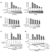
SUV39H1 represses Tax transactivation of HTLV-1 LTR promoter activity. Representative results of luciferase assays using HEK293 and Jurkat cells (left and right panels, respectively) are shown with the mean and standard deviation of triplicate experiments. Below the graphs, results of immunoblot analyses of whole cell lysates are shown to confirm expression of transduced proteins. (a) Dose-dependent repression of Tax transactivation of HTLV-1 LTR by SUV39H1. More than three independent assays were done for each cell line. (b) Effects of SUV39H1 on the basal activities of HTLV-1 LTR. In the absence of Tax, increasing amounts of SUV39H1 expression plasmid was transfected with HTLV-1 Luc. Left and right panels show the results of HEK293 and Jurkat cells, respectively. (c) Absence of repression of Tax transactivation by HMTase negative SUV39H1. Tax expression plasmid was co-transfected with the wild type or HMTase negative mutant SUV39H1 along with the reporter plasmid pHTLV LTR-Luc. Lower two panels show the results of immunoblot analyses to confirm the expression of transduced Tax and SUV39H1 proteins. Antibodies used are indicated on the left. (d) Suppressive activities of SUV39H1 mutants on Tax transactivation of HTLV-1 LTR promoter activity. Fold activation of HTLV LTR promoter activity by Tax is shown with the mean and standard deviation of triplicated experiments. Co-transfected HA-tagged mutant SUV39H1 constructs are indicated below the graph and on the right of lower panels. Structures of these deletion mutants are described in Fig. 2a, upper panel.
Figure 6
Figure 6
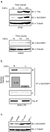
Induction of SUV39H1 expression in JPX9 cells and localization on the HTLV-1 LTR, and endogenous levels of SUV39H1 expression in T cell lines. (a) Top figure: Expression of Tax and SUV39H1 in CdCl2-treated JPX9 cells. Whole cell lysates of JPX9 cells treated by CdCl2 for indicated periods were studied by immunoblot analysis with anti-Tax and anti-SUV39H1 monoclonal antibodies (top and middle panels). The bottom panel shows the immunoblot by anti-tubulin antibody. Bottom figure: Absence of SUV39H1 induction in Jurkat cells by CdCl2 treatment. Whole cell lysates of Jurkat cells treated by CdCl2 for indicated periods were studied by immunoblot analysis with anti-SUV39H1 monoclonal antibody (top panel). The bottom panel shows the immunoblot by anti-tubulin antibody. (b) Results of ChIP assays. Representative photographs of agarose gel electrophoresis of PCR products are shown. Top panel shows results of CdCl2-treated and untreated JPX9LTR clones. The relative intensities of the band measured by NIH Image software are shown in the second panel. The third and bottom panels show the results of negative controls without first antibody and input controls, respectively. (c) SUV39H1 expression in various T cell lines. ATL-derived cell lines (MT-1 and TL-om1) show higher levels of SUV39H1 expression compared with HTLV-1-uninfected cell lines (top panel). TL-om1 and MT-1 are ATL-derived and HTLV-1-infected cell lines. The bottom panel shows the immunoblot by anti-tubulin antibody.

Similar articles

Cited by

References

    1. Poiesz BJ, Ruscetti FW, Gazdar AF, Bunn PA, Minna JD, Gallo RC. Detection and isolation of type C retrovirus particles from fresh and cultured lymphocytes of a patient with cutaneous T-cell lymphoma. Proc Natl Acad Sci U S A. 1980;77:7415–7419. - PMC - PubMed
    1. Yoshida M, Miyoshi I, Hinuma Y. Isolation and characterization of retrovirus from cell lines of human adult T-cell leukemia and its implication in the disease. Proc Natl Acad Sci U S A. 1982;79:2031–2035. - PMC - PubMed
    1. Yamaguchi K, Watanabe T. Human T lymphotropic virus type-I and adult T-cell leukemia in Japan. Int J Hematol. 2002;76 Suppl 2:240–245. - PubMed
    1. Watanabe T. HTLV-1-associated diseases. Int J Hematol. 1997;66:257–278. doi: 10.1016/S0925-5710(97)00077-7. - DOI - PubMed
    1. Koiwa T, Hamano-Usami A, Ishida T, Okayama A, Yamaguchi K, Kamihira S, Watanabe T. 5'-long terminal repeat-selective CpG methylation of latent human T-cell leukemia virus type 1 provirus in vitro and in vivo. J Virol. 2002;76:9389–9397. doi: 10.1128/JVI.76.18.9389-9397.2002. - DOI - PMC - PubMed

Publication types

MeSH terms