. 2006 Feb;116(2):386-94.
doi: 10.1172/JCI26617. Epub 2006 Jan 12.

Independent degeneration of photoreceptors and retinal pigment epithelium in conditional knockout mouse models of choroideremia

Affiliations

Independent degeneration of photoreceptors and retinal pigment epithelium in conditional knockout mouse models of choroideremia

Tanya Tolmachova et al. J Clin Invest. 2006 Feb.

Abstract

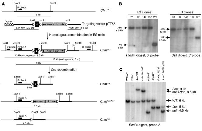
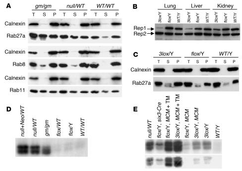
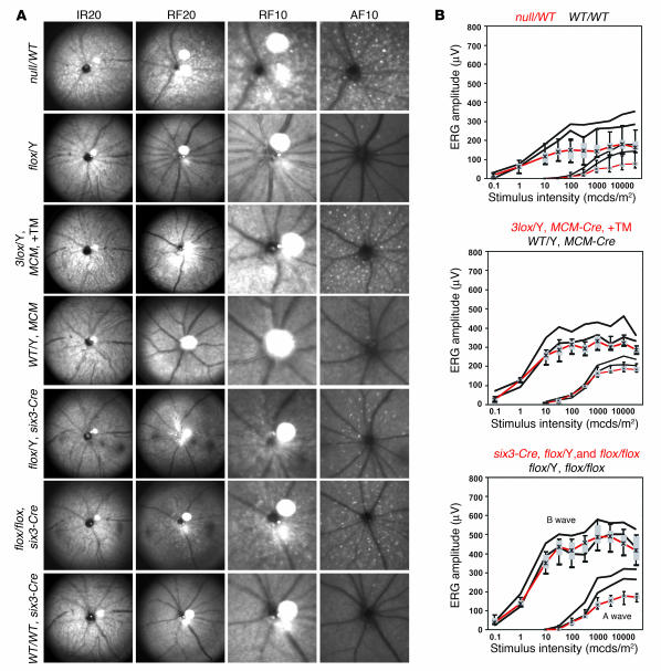
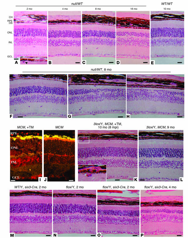
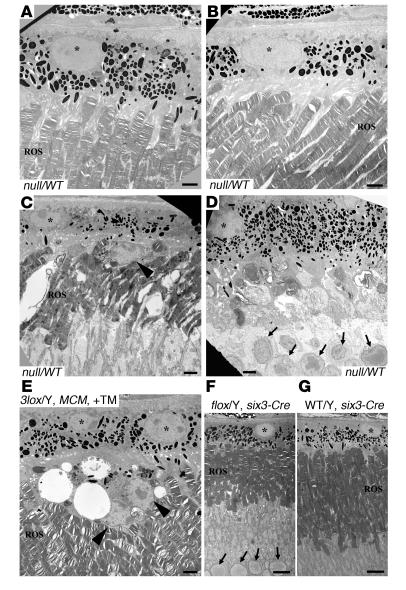
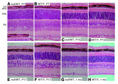
Choroideremia (CHM) is an X-linked degeneration of the retinal pigment epithelium (RPE), photoreceptors, and choroid, caused by loss of function of the CHM/REP1 gene. REP1 is involved in lipid modification (prenylation) of Rab GTPases, key regulators of intracellular vesicular transport and organelle dynamics. To study the pathogenesis of CHM and to develop a model for assessing gene therapy, we have created a conditional mouse knockout of the Chm gene. Heterozygous-null females exhibit characteristic hallmarks of CHM: progressive degeneration of the photoreceptors, patchy depigmentation of the RPE, and Rab prenylation defects. Using tamoxifen-inducible and tissue-specific Cre expression in combination with floxed Chm alleles, we show that CHM pathogenesis involves independently triggered degeneration of photoreceptors and the RPE, associated with different subsets of defective Rabs.


Figures

Figure 1
Figure 1
Generation of mice carrying the conditional and KO Chm alleles. (A) Targeting vector pTT55 carrying 3 loxP sites, a neomycin and spectinomycin resistance cassette (Neor, Spr), and 2 homology arms was used to generate the Chm3lox allele in GSI-1 ES cells by homologous recombination. Diagnostic HindIII and SstI restriction sites and corresponding 3′ and 5′ probes were used to identify correctly targeted ES clones. Cre-mediated recombination between the 3 loxP sites within the Chm3lox allele resulted in 3 possible alleles: Chmflox, Chmnull+Neo, and Chmnull, which were distinguished by Southern blot analysis using EcoRI digestion and probe A. (B) Results of Southern blot analysis using HindIII digestion and the 3′ probe, and SstI digestion and the 5′ probe, are shown for 4 correctly targeted ES clones (78, 92, 147, and 197) and a wild-type ES clone. (C) Results of Southern blot analysis using EcoRI digestion and probe A. The Chm alleles of the mice are indicated above the lanes. “MCM” indicates mice carrying the MerCreMer transgene. “+TM” indicates treatment with TM.
Figure 2
Figure 2
Analysis of Rep1 protein amount and function. (A) Total protein lysates from stomach of gunmetal (gm/gm), Chmnull/WT, and ChmWT/WT mice were subjected to subcellular fractionation, and total (T), soluble (S), and pellet (P) fractions were immunoblotted and probed with anti-Rab27a antibody 4B12, anti-Rab8 antibody, and anti-Rab11 antibody as indicated. Calnexin was used as a control for subcellular fractionation. (B) Immunoblot of total protein lysates from lung, liver, and kidney of Chm3lox/Y, Chmflox/Y, and ChmWT/Y mice probed with anti-Rep antibody J905, which recognizes both Rep1 and Rep2 proteins. (C) Total protein lysates from large intestines of Chm3lox/Y, Chmflox/Y, and ChmWT/Y mice were subjected to subcellular fractionation, and total (T), soluble (S), and pellet (P) fractions were immunoblotted and probed with anti-Rab27a antibody 4B12. Calnexin was used as a control for subcellular fractionation. (D and E) In vitro prenylation was carried out on the soluble fraction of total protein lysates from large intestines (D) and eyes (E). The Chm alleles are indicated above the lanes. “MCM” indicates mice carrying the MerCreMer transgene. “+TM” indicates treatment with TM. The radiogram underneath shows a shorter exposure to film.
Figure 3
Figure 3
Ophthalmic and electroretinographic analysis of Chm mutant mouse eyes. (A) Retinal SLO images from representative Chm mutant animals. The Chm alleles, MerCreMer (MCM) transgene, six3-Cre transgene, and TM induction (+TM) are indicated to the left of each set of panels. An equipment setting of 20° was used for a full view and of 10° for a more detailed view. The wavelengths shown are infrared (890 nm and 20°, IR20), green (514 nm and 20° or 10°, RF20 and RF10), and blue autofluorescence (488 nm with barrier at 500 nm and 10°, AF10). (B) Electroretinographic analysis showing a and b waves for the indicated strains of mice. Box and whisker plot of mutant mouse data (red) (whiskers 5% and 95% quantile, box 25–75% quantile, asterisk median) in comparison with respective control mouse data (black) (5% and 95% quantiles indicated by the lower and upper black lines, respectively, delimiting a 90% normal range). mcds, millicandela seconds.
Figure 4
Figure 4
Histological analysis of sections of eyes of Chm mutant mice. (AH) Chmnull/WT mice at 2 (A), 4 (B), 8 (C and FH), and 16 (D) months, and ChmWT/WT mice at 10 months (E). (I and J) Detection of Cre protein (red) in TM-induced (+TM) (I) and non–TM-induced (J) mice carrying the MerCreMer transgene (MCM) with a Cre-specific antibody. (K and L) Histological analysis of TM-induced (+TM) (K) and non–TM-induced (L) Chm3lox/Y mice carrying the MerCreMer transgene (MCM) at 9–10 months. Analysis was carried out 8 months after TM injection (8 mpi). (MP) Two-month-old ChmWT/Y (M) and Chmflox/Y (N) mice, and Chmflox/Y six3-Cre+ mice at 2 (O) and 4 (P) months. CH, choroid; RPE, retinal pigment epithelium; OS, photoreceptor outer segments; ONL, outer nuclear layer; INL, inner nuclear layer; GCL, ganglion cell layer. Scale bars: 20 μm. Magnification, ×230 (A, inset), ×450 (K, inset).
Figure 5
Figure 5
Electron microscopic analysis of Chm mutant retinas. (AD) Different sections from the eye of a 9-month-old Chmnull/WT female mouse. (E) A TM-induced Chm3lox/Y MerCreMer mouse. (F) A Chmflox/Y six3-Cre mouse. (G) A ChmWT/Y six3-Cre mouse. RPE nuclei are marked by asterisks. Abnormal cells above the RPE are shown by arrowheads in C and E. Nuclei of cells from the ONL are marked by arrows in D and F. Scale bars: 2 μm (AE) or 7 μm (F and G).
Figure 6
Figure 6
Histological analysis of Chm mutant retinas during development. Sections from Chmnull/WT and ChmWT/Y littermates at P7, P15, and P17 and at 1 month as indicated for each panel. Magnification, ×125 (AH), ×210 (A, inset).

References

    1. Pacione LR, Szego MJ, Ikeda S, Nishina PM, McInnes RR. Progress toward understanding the genetic and biochemical mechanisms of inherited photoreceptor degenerations. Annu. Rev. Neurosci. 2003;26:657–700. - PubMed
    1. Stone EM, Sheffield VC, Hageman GS. Molecular genetics of age-related macular degeneration. Hum. Mol. Genet. 2001;10:2285–2292. - PubMed
    1. Strauss O. The retinal pigment epithelium in visual function. Physiol. Rev. 2005;85:845–881. - PubMed
    1. Heckenlively, J.R., and Bird, A.J. 1988. Choroideremia. In Retinitis pigmentosa. J.R. Heckenlively, editor. Lippincott Williams & Wilkins. Philadelphia, Pennsylvania, USA. 176–187.
    1. Cremers, F.P.M. 1995. Choroideremia. In The metabolic and molecular bases of inherited disease. C.B. Scriver, A.L. Beaudet, W.S. Sly, and D. Valle, editors. McGraw-Hill. New York, New York, USA. 4311–4324.

Publication types

MeSH terms

Substances