. 2006 Feb;116(2):414-21.
doi: 10.1172/JCI26631. Epub 2006 Jan 12.

Transcription factor T-bet regulates inflammatory arthritis through its function in dendritic cells

Affiliations

Transcription factor T-bet regulates inflammatory arthritis through its function in dendritic cells

Jingsong Wang et al. J Clin Invest. 2006 Feb.

Abstract

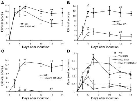
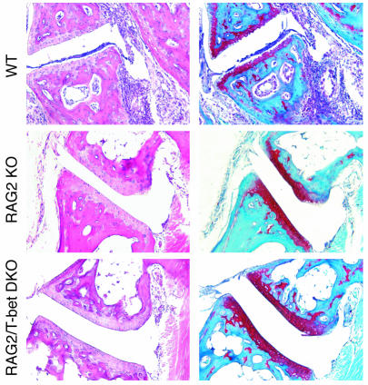
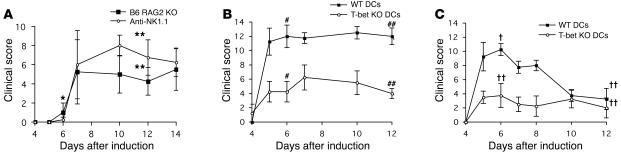
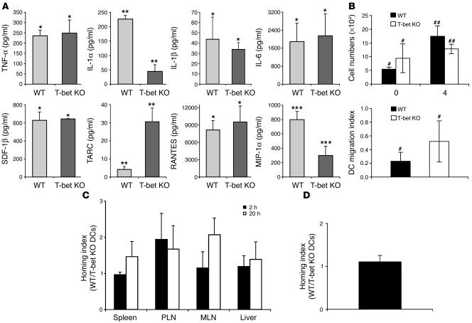
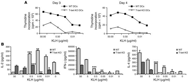
The transcription factor T-bet (Tbx21) plays a major role in adaptive immunity and is required for optimal IFN-gamma production by DCs. Here we demonstrate an essential function for T-bet in DCs in controlling inflammatory arthritis. We show that collagen antibody-induced arthritis (CAIA), a model of human RA, is a bipartite disease characterized by an early innate immune system component intact in RAG2 mice and a later adaptive immune system phase. Mice lacking T-bet had markedly reduced joint inflammation at both early and late time points and RAG2T-bet double-deficient mice were essentially resistant to disease. Remarkably, adoptive transfer of T-bet-expressing DCs reconstituted inflammation in a T-bet deficient and T-bet/RAG2-deficient milieu. T-bet regulates the production of proinflammatory cytokine IL-1alpha and chemokines macrophage inflammatory protein-1alpha (MIP-1alpha) and thymus- and activation-related chemokine (TARC) by DCs. Further, T-bet expression in DCs is required for T helper cell activation. We conclude that T-bet plays a vital function in DCs that links innate and adaptive immunity to regulate inflammatory responses. T-bet provides an attractive new target for the development of novel therapeutics for inflammatory arthritis.


Figures

Figure 1
Figure 1
T-bet is expressed in human rheumatoid synovium. Immunohistochemical analysis of synovial tissue from 7 patients with established RA who fulfilled American College of Rheumatology criteria for the diagnosis of RA. (A) H&E staining. (BF) Representative immunostaining of inflammatory synovium for (B) T-bet, (C) CD3 (T cells), (D) CD20 (B cells), (E) CD68 (macrophages), and (F) S100 (DCs).
Figure 2
Figure 2
CAIA can be induced in both WT and RAG2–/– mice and is diminished in the absence of T-bet. Seven-week-old BALB/c mice (WT) and (A) RAG2–/– mice on BALB/c background (RAG2 KO), (B) T-bet–/– mice (T-bet KO), and (C) RAG2–/–T-bet–/– DKO mice (RAG2/T-bet DKO) were subjected to arthritis induction. Clinical signs of inflammation (AC) and paw swelling (D) were scored and plotted at different time points as indicated. The values are expressed as mean ± SEM. Each group consists of 4 mice. The results shown are representative of 3 to 5 separate experiments. Unpaired Student’s t tests were performed on days 6 and 12 after arthritis induction. *P > 0.3; **P < 0.05; #P < 0.02; ##P < 0.002; P < 0.001; ††P < 0.001.
Figure 3
Figure 3
Histologic analysis of CAIA. Tissue sections obtained from paw joints of WT (upper), RAG2–/– (middle), and RAG2–/–T-bet–/– DKO (lower) mice on day 14 after arthritis induction were stained with H&E (left panels) and safranin O red (right panels). Inflammatory cell accumulation in synovium and loss of safranin O red staining were abundant in WT but not in RAG2–/– or RAG2–/–T-bet–/– DKO mice. The results shown are representative images from 3 to 6 independent experiments with 4 mice per group in each experiment.
Figure 4
Figure 4
Adoptive transfer of WT but not T-bet KO DCs restores inflammatory arthritis. Clinical signs of inflammation were scored and plotted at different time points as indicated. Individual cell components of the innate immune system were evaluated for their role in the development of arthritis. (A) RAG2–/– on C57BL/6 background (B6 RAG2 KO) and the same strain of mice depleted of NK cells in vivo by repeated injection of anti-NK1.1 (anti-NK1.1). (B) T-bet KO mice and (C) RAG2–/–T-bet–/– DKO mice received 0.3 × 10⁶ purified WT or T-bet KO splenic DCs. Unpaired Student’s t tests were performed on days 6 and 12 after arthritis induction. *P > 0.5; **P > 0.3; #P < 0.02; ##P < 0.001; P < 0.003; ††P > 0.2.
Figure 5
Figure 5
T-bet controls the production of inflammatory cytokines and chemokines in DCs but does not regulate DC migration and life span in vivo. (A) Cytokine and chemokine levels in DC culture supernatants were measured with SearchLight high dynamic range imaging and analysis system. Culture supernatants were collected from T-bet–/– or WT DCs after in vitro culture with LPS for 20 hours. Results are from 3 independent experiments with DCs pooled from 8 mice per group in each experiment. SDF-1β, stromal cell–derived factor-1β. (B) CD11c+ DC counts in knee joint synovium of WT and T-bet–/– mice at days 0 and 4 after arthritis induction (top panel). On day 4 after arthritis induction, ratios of CD11c+ DCs in popliteal LNs and knee joint synovium were calculated as migration index in WT and T-bet–/– mice (bottom panel). *P > 0.05; **P < 0.005; ***P < 0.03; #P > 0.05; ##P > 0.05. (C) Homing ratio (WT/T-bet KO) measurement of i.v.-injected DCs in WT and T-bet KO mice. PLN, peripheral LNs; MLN, mesenteric LNs. Results are from 9 independent experiments. (D) Homing ratio (WT/T-bet KO) of footpad-injected DCs in popliteal LNs at 20 hours after injection. Results are from 6 independent experiments.
Figure 6
Figure 6
T-bet–/– DCs are defective in activating T cells in vivo. (A) LN cell proliferation to KLH on day 3 of popliteal LN cells harvested from WT mice primed with KLH-pulsed T-bet–/– or WT DCs 3 and 4 days earlier. The results shown are representative of 3 independent experiments. (B) Cytokine production from LN cells primed with KLH-loaded WT and T-bet KO DCs and cultured in the presence of various concentrations (from 0.01 to 50 μg/ml) of KLH at the time points indicated. *P < 0.05; **P < 0.01; ***P < 0.05.

References

    1. Stastny P. Association of the B-cell alloantigen DRw4 with rheumatoid arthritis. N. Engl. J. Med. 1978;298:869–871. - PubMed
    1. Firestein GS. Evolving concepts of rheumatoid arthritis. Nature. 2003;423:356–361. - PubMed
    1. Plows D, Kontogeorgos G, Kollias G. Mice lacking mature T and B lymphocytes develop arthritic lesions after immunization with type II collagen. J. Immunol. 1999;162:1018–1023. - PubMed
    1. Santiago-Schwarz F, Anand P, Liu S, Carsons SE. Dendritic cells (DCs) in rheumatoid arthritis (RA): progenitor cells and soluble factors contained in RA synovial fluid yield a subset of myeloid DCs that preferentially activate Th1 inflammatory-type responses. J. Immunol. 2001;167:1758–1768. - PubMed
    1. Matsumoto I, Staub A, Benoist C, Mathis D. Arthritis provoked by linked T and B cell recognition of a glycolytic enzyme. Science. 1999;286:1732–1735. - PubMed

Publication types

MeSH terms