

Relatively small increases in the steady-state levels of nucleobase deamination products in DNA from human TK6 cells exposed to toxic levels of nitric oxide
- PMID: 16411656
- PMCID: PMC2515361
- DOI: 10.1021/tx050252j
Relatively small increases in the steady-state levels of nucleobase deamination products in DNA from human TK6 cells exposed to toxic levels of nitric oxide
Abstract
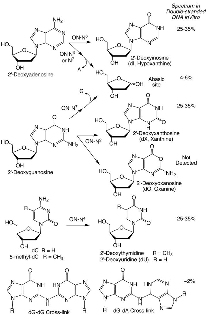
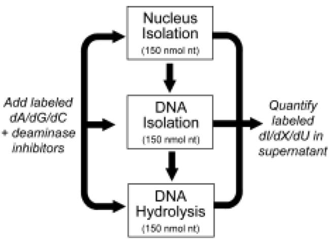
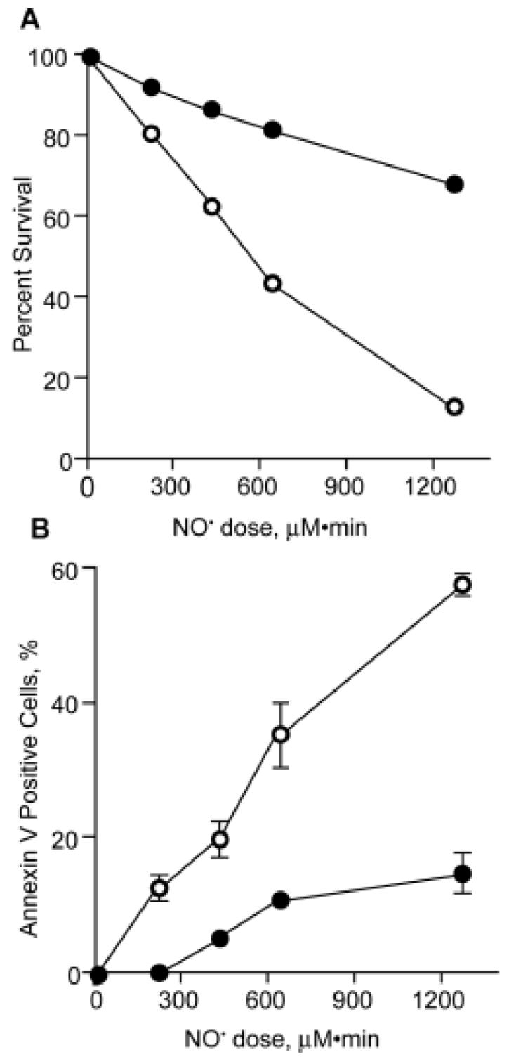
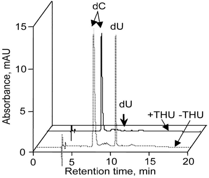
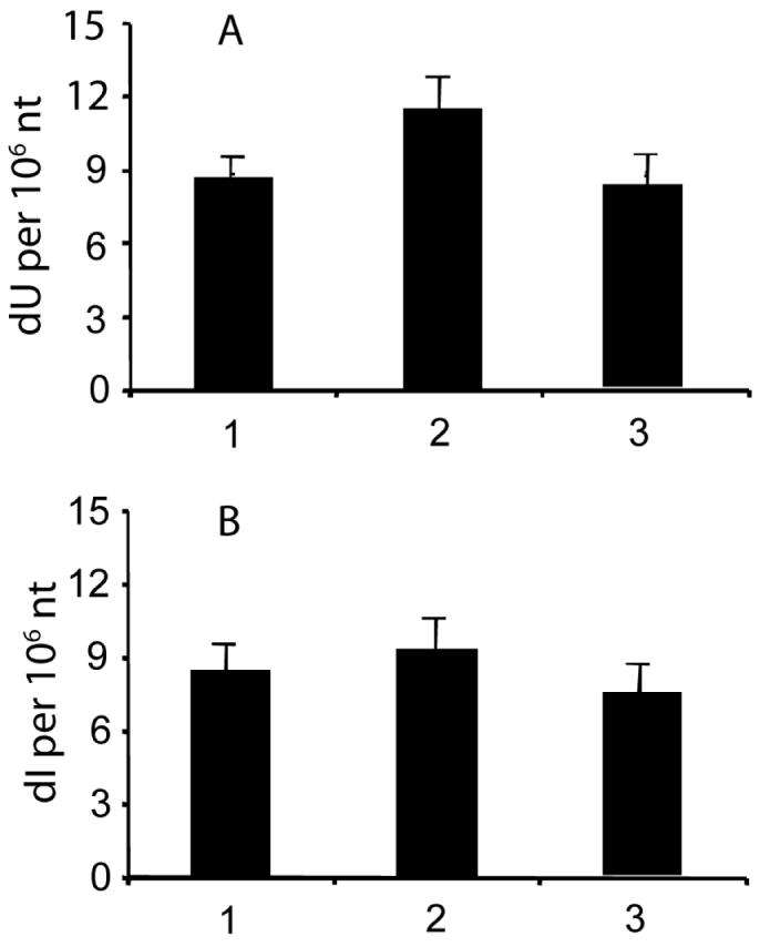
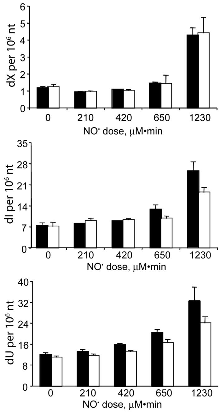
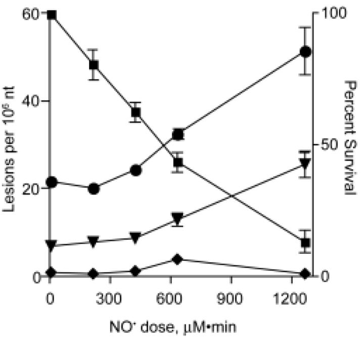
Nitric oxide (NO) is a physiologically important molecule that has been implicated in the pathophysiology of diseases associated with chronic inflammation, such as cancer. While the complicated chemistry of NO-mediated genotoxicity has been extensively study in vitro, neither the spectrum of DNA lesions nor their consequences in vivo have been rigorously defined. We have approached this problem by exposing human TK6 lymphoblastoid cells to controlled steady-state concentrations of 1.75 or 0.65 microM NO along with 186 microM O2 in a recently developed reactor that avoids the anomalous gas-phase chemistry of NO and approximates the conditions at sites of inflammation in tissues. The resulting spectrum of nucleobase deamination products was defined using a recently developed liquid chromatography/mass spectrometry (LC/MS) method, and the results were correlated with cytotoxicity and apoptosis. A series of control experiments revealed the necessity of using dC and dA deaminase inhibitors to avoid adventitious formation of 2'-deoxyuridine (dU) and 2'-deoxyinosine (dI), respectively, during DNA isolation and processing. Exposure of TK6 cells to 1.75 microM NO and 186 microM O2 for 12 h (1260 microM x min dose) resulted in 32% loss of cell viability measured immediately after exposure and 87% cytotoxicity after a 24 h recovery period. The same exposure resulted in 3.5-, 3.8-, and 4.1-fold increases in dX, dI, and dU, respectively, to reach the following levels: dX, 7 (+/- 1) per 10(6) nt; dI, 25 (+/- 2.1) per 10(6) nt; and dU, 40 (+/- 3.8) per 10(6) nt. dO was not detected above the limit of detection of 6 lesions per 10(7) nt in 50 microg of DNA. A 12 h exposure to 0.65 microM NO and 190 microM O2 (468 microM x min dose) caused 1.7-, 1.8-, and 2.0-fold increases in dX, dI, and dU, respectively, accompanied by a approximately 15% (+/- 3.6) reduction in cell viability immediately after exposure. Again, dO was not detected. These results reveal modest increases in the steady-state levels of DNA deamination products in cells exposed to relatively cytotoxic levels of NO. This could result from limited nitrosative chemistry in nuclear DNA in cells exposed to NO or high levels of formation balanced by rapid repair of nucleobase deamination lesions in DNA.
Figures
References
-
- Moncada S, Palmer RM, Higgs EA. Nitric oxide: physiology, pathophysiology, and pharmacology. Pharmacol. Rev. 1991;43:109–142. - PubMed
-
- Nathan C, Xie QW. Nitric oxide synthases: roles, tolls, and controls. Cell. 1994;78:915–918. - PubMed
-
- Schmidt HHHW, Walter U. NO at work. Cell. 1994;78:919–925. - PubMed
-
- Ohshima H. Genetic and epigenetic damage induced by reactive nitrogen species: implications in carcinogenesis. Toxicol. Lett. 2003;140-141:99–104. - PubMed
-
- Dedon PC, Tannenbaum SR. Reactive nitrogen species in the chemical biology of inflammation. Arch. Biochem. Biophys. 2004;423:12–22. - PubMed
Publication types
MeSH terms
Substances
Grants and funding
LinkOut - more resources
Full Text Sources
Other Literature Sources
Research Materials
