. 2006 Jan 24;45(3):746-54.
doi: 10.1021/bi050870z.

Photolabeling of cardiolipin binding subunits within bovine heart cytochrome c oxidase

Affiliations

Photolabeling of cardiolipin binding subunits within bovine heart cytochrome c oxidase

Erik Sedlák et al. Biochemistry. .

Abstract

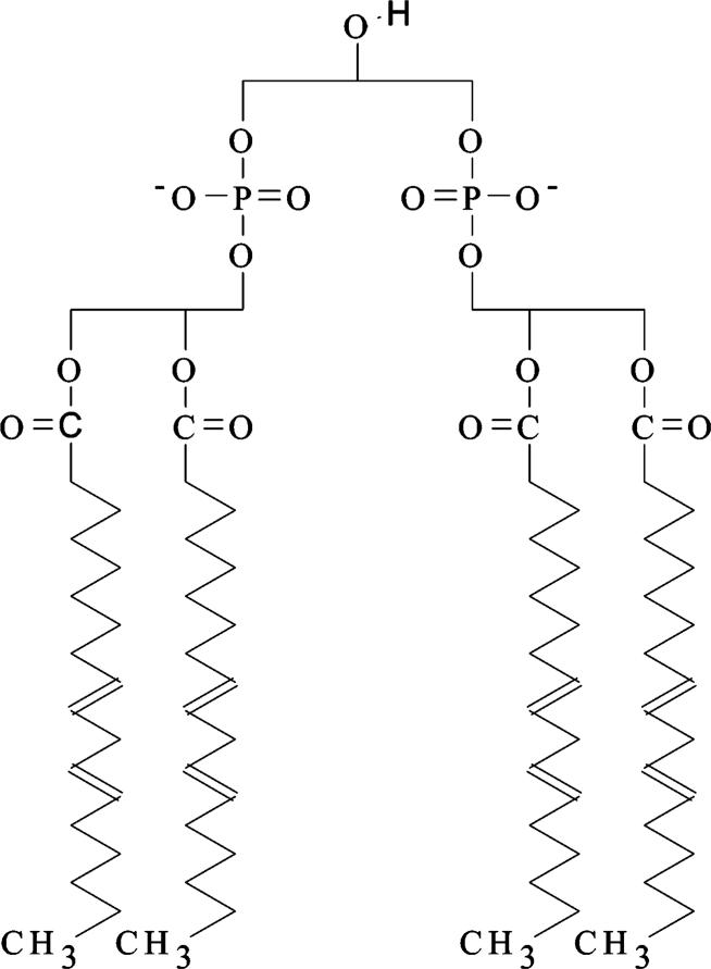
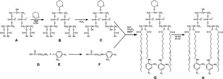
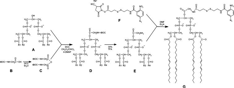
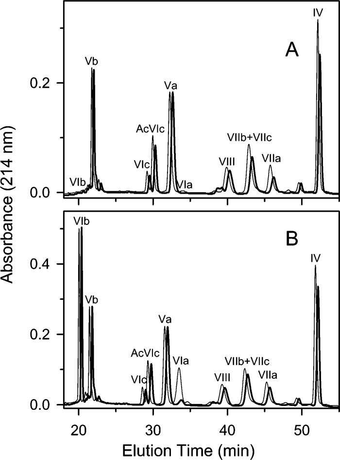
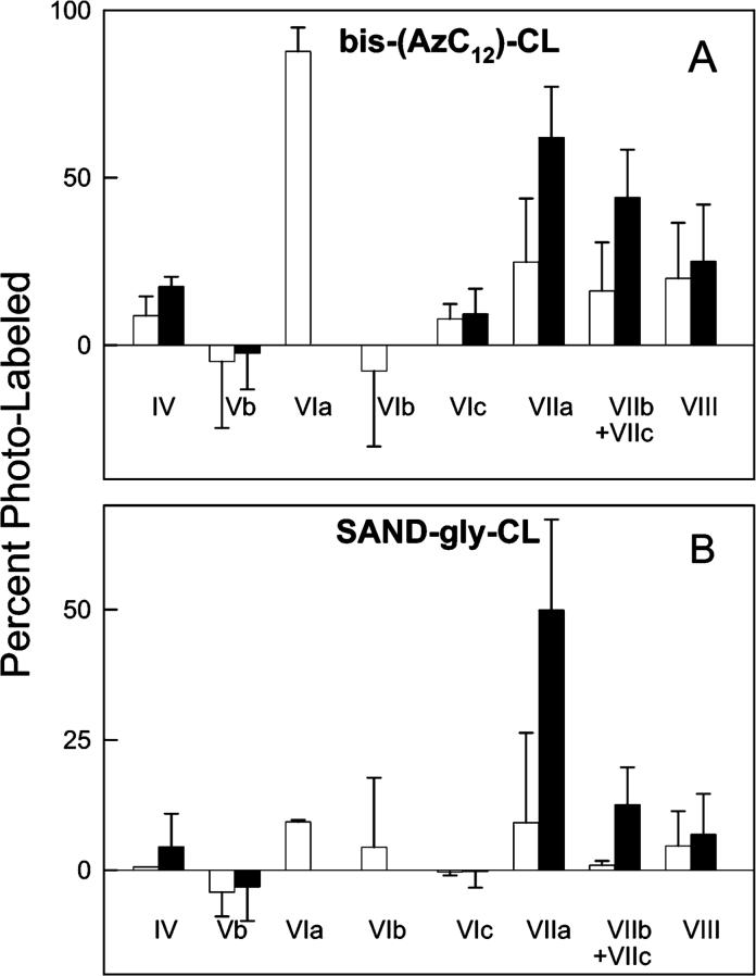
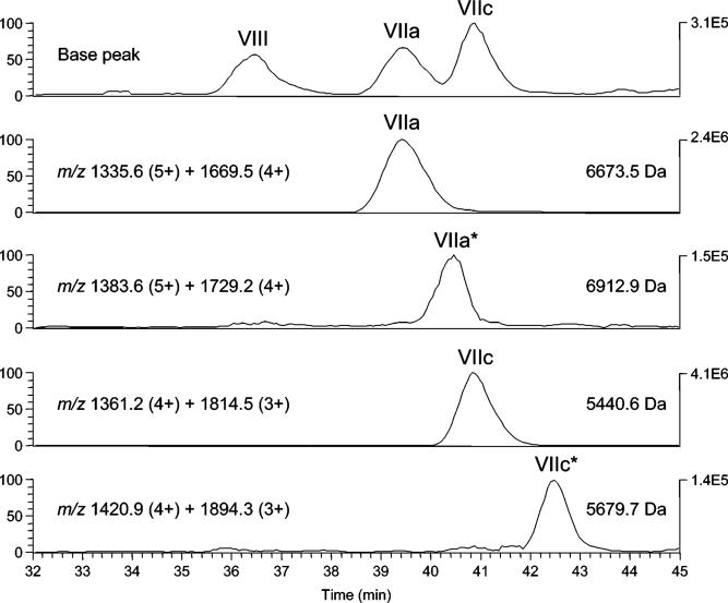
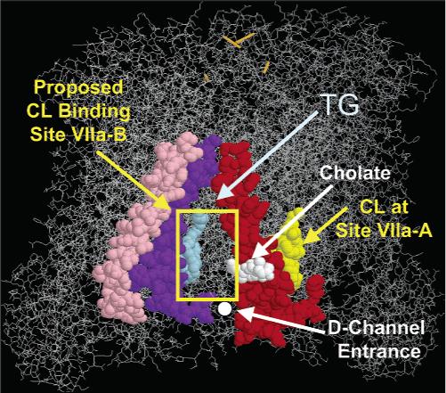
Subunits located near the cardiolipin binding sites of bovine heart cytochrome c oxidase (CcO) were identified by photolabeling with arylazido-cardiolipin analogues and detecting labeled subunits by reversed-phase HPLC and HPLC-electrospray ionization mass spectrometry. Two arylazido-containing cardiolipin analogues were synthesized: (1) 2-SAND-gly-CL with a nitrophenylazido group attached to the polar headgroup of cardiolipin (CL) via a linker containing a cleavable disulfide; (2) 2',2''-bis-(AzC12)-CL with two of the four fatty acid tails of cardiolipin replaced by 12-(N-4-azido-2-nitrophenyl) aminododecanoic acid. Both arylazido-CL derivatives were used to map the cardiolipin binding sites within two types of detergent-solubilized CcO: (1) intact 13-subunit CL-containing CcO (three to four molecules of endogenous CL remain bound per CcO monomer); (2) 11-subunit CL-free CcO (subunits VIa and VIb are missing because they dissociate during CL removal). Upon the basis of these photolabeling studies, we conclude that (1) subunits VIIa, VIIc, and possibly VIII are located near the two high-affinity cardiolipin binding sites, which are present in either form of CcO, and (2) subunit VIa is located adjacent to the lower affinity cardiolipin binding site, which is only present in the 13-subunit form of CcO. These data are consistent with the recent CcO crystal structure in which one cardiolipin is located near subunit VIIa and a second is located near subunit VIa (PDB ID code referenced in Tomitake, T. et al. (2003) Proc. Natl. Acad. Sci. U.S.A. 100, 15304-15309). However, we propose that a third cardiolipin is bound between subunits VIIa and VIIc near the entrance to the D-channel. Cardiolipin bound at this location could potentially function as a proton antenna to facilitate proton entry into the D-channel. If true, it would explain the CcO requirement of bound cardiolipin for full electron transport activity.


Figures

Figure 1
Figure 1
Cardiolipin (diphosphatidyl-glycerol).
Figure 2
Figure 2
Synthesis of bis-(AzC12)-cardiolipin: (A) cardiolipin; (B) THP-CL; (C) dilyso-THP-CL; (D) 12-aminododecanoic acid; (E) 4-fluoro-3-nitrophenyl azide; (F) ANPA; (G) bis-(AzC12)-THP-CL; (H) bis-(AzC12)-CL.
Figure 3
Figure 3
Synthesis of SAND-gly-cardiolipin: (A) cardiolipin; (B) t-BOC-glycine; (C) t-BOC-glycine anhydride; (D) t-BOC-glycyl-CL; (E) 2-glycyl-CL; (F) SAND; (G) SAND-gly-CL.
Figure 4
Figure 4
Detection of bis-(AzC12)-CL photolabeling of cytochrome c oxidase subunits by RP-HPLC subunit analysis. Percent photolabeling of each subunit was indirectly determined by quantifying the percent of each subunit that was unmodified after normalizing the two chromatograms to make the percent yield of subunit Va equal. Panel A shows RP-HPLC analysis of CL-free CcO subunits before (thin line) and after (thick line) photoactivation of bound bis-(AzC12)-CL. In this experiment, the yields of subunits IV, VIIa, VIIb/VIIc, and VIII decreased by 19%, 47%, 39%, and 12%, respectively. Panel B shows RP-HPLC analysis of CL-containing CcO subunits before (thin line) and after (thick line) photoactivation of bound bis-(AzC12)-CL. In this experiment, the yields of subunits IV, VIIa, VIIb/VIIc, and VIII decreased by 9%, 21%, 12%, and 17%, respectively. In both panels, the chromatogram for photolabeled CcO (thick line) has been offset by 0.6 min to improve clarity and to facilitate its comparison with the chromatogram for the unlabeled CcO control (thin line). In each analysis, 100 μg (0.5 nmol) of CcO was loaded on the RP-column, and the subunit content was quantitatively determined according to Liu et al. (30).
Figure 5
Figure 5
Photolabeling of cytochrome c oxidase by bis-(AzC12)-CL or SAND-gly-CL. Panel A shows photolabeling of CL-free (filled bars) and CL-containing CcO (open bars) by bis-(AzC12)-CL. Panel B shows photolabeling of CL-free (filled bars) and CL-containing CcO (open bars) by SAND-gly-CL. Percent photolabeling was calculated from the decreased area of the RP-HPLC elution peak for enzyme that had been reconstituted with either bis-(AzC12)-CL or SAND-gly-CL, purified by HiTrap Q ion exchange chromatography to remove free ligand, and exposed or not exposed to intense visible light. Average values and standard deviations were calculated from three separate paired experiments. The percent labeling of subunit VIa is based upon the total decreased HPLC yield of subunit VIa plus Ac-VIa. In each analysis, 100 μg (0.5 nmol) of CcO was loaded on the RP-column, and the subunit content was quantitatively determined according to Liu et al. (30).
Figure 6
Figure 6
RP-HPLC–ESI/MS analysis of ANBET-modified CcO subunits. Eleven-subunit CL-free CcO was photolabeled with SAND-gly-CL, treated with dithiothreitol, and analyzed by RP-HPLC–ESI/MS, as described in Methods. The top panel shows the base peak chromatogram (plot of the intensity of the most abundant ion in each spectrum) for components eluting in the time range of interest (32−45 min). The lower four panels contain selected ion retrieval traces generated by the sum of the intensities of the indicated multiply charged ions. The resulting molecular mass of each species is indicated on the right side of each panel. Elution peaks labeled VIIa* and VIIc* are the ANBET-modified subunits, that is, +239 Da compared to unlabeled subunit VIIa and VIIc.
Figure 7
Figure 7
Cardiolipin binding sites near subunit VIIa. Two CL binding sites are proposed to be located adjacent to subunit VIIa. CL bound at the first site (site VIIa-A) is visible in the three-dimensional crystal structure of CcO. A second CL binding site (site VIIa-B) is proposed to be located between subunits VIIa (red) and VIIc (purple), in contact with subunit I (wireframe) and close to subunit VIII (pink). The CL headgroup would be bound adjacent to the entrance to the D-channel. A single CL (yellow), cholate (white), and triglyceride (TG, light blue) are located close to these subunits within the crystal structure. The figure was prepared using the atomic coordinates of CcO in the Protein Data Bank, PDB ID code 1v54 (14).

References

    1. Kadenbach B, Jarausch J, Hartmann R, Merle P. Separtation of Mammalian Cytochrome c Oxidase into 13 Polypeptides by a Sodium Dodecyl Sulfate-Gel Electrophoresis Procedure. Anal. Biochem. 1983;129:517–521. - PubMed
    1. Ioannou PV, Golding BT. Cardiolipins: Their Chemistry and Biochemistry. J. Lipid Res. 1979;17:279–318. - PubMed
    1. Hoch FL. Cardiolipins and Biomembrane Function. Biochim. Biophys. Acta. 1992;1113:71–133. - PubMed
    1. Schlame M, Rua D, Greenberg ML. The Biosynthesis and Functional Role of Cardiolipin. Prog. Lipid Res. 2000;39:257–288. - PubMed
    1. Robinson NC. Functional Binding of Cardiolipin to Cytochromec Oxidase. J. Bioenerg. Biomembr. 1993;25:153–163. - PubMed

Publication types