. 2006 May 1;395(3):457-62.
doi: 10.1042/BJ20051936.

Crystal structure of inactivated Thermotoga maritima invertase in complex with the trisaccharide substrate raffinose

Affiliations

Crystal structure of inactivated Thermotoga maritima invertase in complex with the trisaccharide substrate raffinose

François Alberto et al. Biochem J. .

Abstract

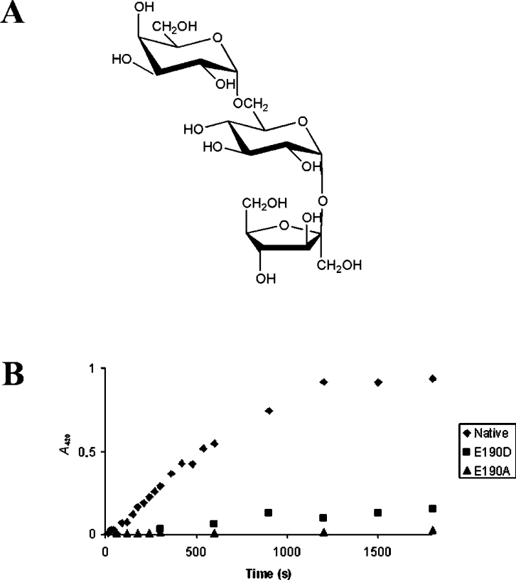
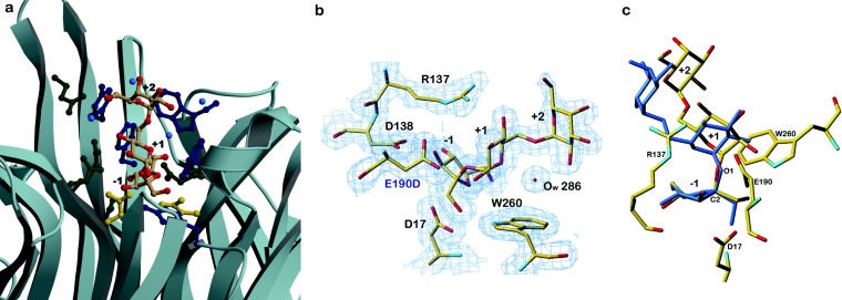
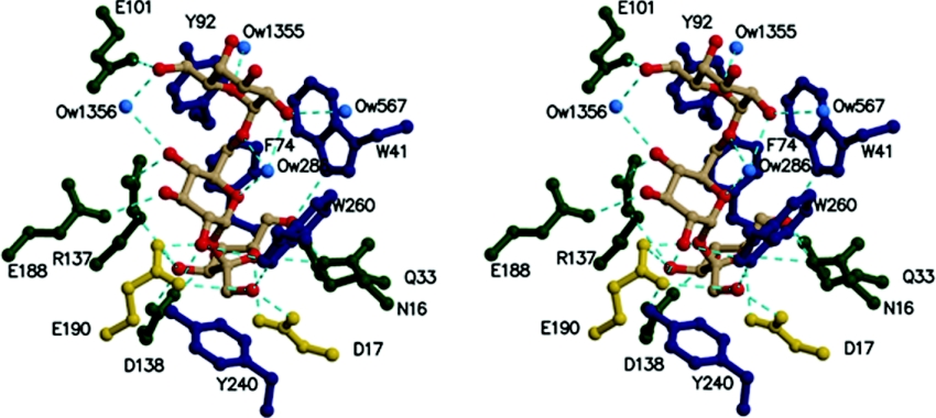
Thermotoga maritima invertase (beta-fructosidase), a member of the glycoside hydrolase family GH-32, readily releases beta-D-fructose from sucrose, raffinose and fructan polymers such as inulin. These carbohydrates represent major carbon and energy sources for prokaryotes and eukaryotes. The invertase cleaves beta-fructopyranosidic linkages by a double-displacement mechanism, which involves a nucleophilic aspartate and a catalytic glutamic acid acting as a general acid/base. The three-dimensional structure of invertase shows a bimodular enzyme with a five bladed beta-propeller catalytic domain linked to a beta-sandwich of unknown function. In the present study we report the crystal structure of the inactivated invertase in interaction with the natural substrate molecule alpha-D-galactopyranosyl-(1,6)-alpha-D-glucopyranosyl-beta-D-fructofuranoside (raffinose) at 1.87 A (1 A=0.1 nm) resolution. The structural analysis of the complex reveals the presence of three binding-subsites, which explains why T. maritima invertase exhibits a higher affinity for raffinose than sucrose, but a lower catalytic efficiency with raffinose as substrate than with sucrose.


Figures

Figure 1
Figure 1. (A) Schematic representation of the substrate molecule α-D-galactopyranosyl-(1,6)-α-D-glucopyranosyl-β-D-fructofuranoside (raffinose). (B) Comparison of the activity between native invertase (◆) with mutants’ inv-E190D (■) and inv-E190A (▲)
Glucose release after sucrose hydrolysis was assayed by the colorimetric reaction of the reducing ends with ferricyanide. The activity was monitored by the decrease in A420 as a function of time (scale in seconds).
Figure 2
Figure 2. Ribbon representation of the superimposition of the three three-dimensional structures of GH32 family enzymes
Tm invertase is coloured in red, Ci fructan 1-exohydrolase in blue and Aa inolinase in yellow. The arrows highlight specific loop insertions that most probably are responsible for modifying the substrate specificities of the three enzymes. Figures 2, 3a and 4 were produced with Molscript [30,31] and rendered with Raster3D.
Figure 3
Figure 3. Localization and structural arrangement of raffinose bound in the active-site pocket in Tm invertase
(a) Ribbon representation of the active-site pocket in Tm invertase. The raffinose molecule trapped in the active site is shown in coloured atoms as a stick-representation. (Oxygen atoms are coloured in red). Catalytic residues are shown in gold, aromatic residues in dark blue and other residues in green. Blue balls are water molecules. The substrate-binding subsites are annotated from −1 to +2. (b) Electron density calculated after the final refinement step for the Tm invertase inv-E190D in complex. The Fourier map (2FoFc) is contoured at 1σ showing the trapped substrate molecule present in the active site of inv-E190D. (c) Stick representation of raffinose in complex with Tm inv-E190D (standard atom type colours) superimposed on to the crystal structure of raffinose pentahydrate (in blue). Figures 3(b) and 3(c) were prepared with TURBO-FRODO [19].
Figure 4
Figure 4. Stereographic view of the catalytic site of invertase in complex with raffinose
Catalytic residues are shown in gold, aromatic residues in dark blue and other residues in green. Hydrogen bonds are in cyan. Ow designates water molecules.

References

    1. Ritsema T., Smeekens S. Fructans: beneficial for plants and humans. Curr. Opin. Plant Biol. 2003;6:223–230. - PubMed
    1. Sturm A., Tang G. Q. The sucrose-cleaving enzymes of plants are crucial for development, growth and carbon partitioning. Trends Plant Sci. 1999;4:401–407. - PubMed
    1. Sturm A. Invertases. Primary structures, functions, and roles in plant development and sucrose partitioning. Plant Physiol. 1999;121:1–8. - PMC - PubMed
    1. Henrissat B. A classification of glycosyl hydrolases based on amino acid sequence similarities. Biochem. J. 1991;280:309–316. - PMC - PubMed
    1. Koshland D. E., Jr, Stein S. S. Correlation of bond breaking with enzyme specificity; cleavage point of invertase. J. Biol. Chem. 1954;208:139–148. - PubMed

Publication types

MeSH terms