Comparative Study
. 2006 Apr 15;395(2):417-26.
doi: 10.1042/BJ20051321.

The CD38-independent ADP-ribosyl cyclase from mouse brain synaptosomes: a comparative study of neonate and adult brain

Affiliations
Comparative Study

The CD38-independent ADP-ribosyl cyclase from mouse brain synaptosomes: a comparative study of neonate and adult brain

Claire Ceni et al. Biochem J. .

Abstract

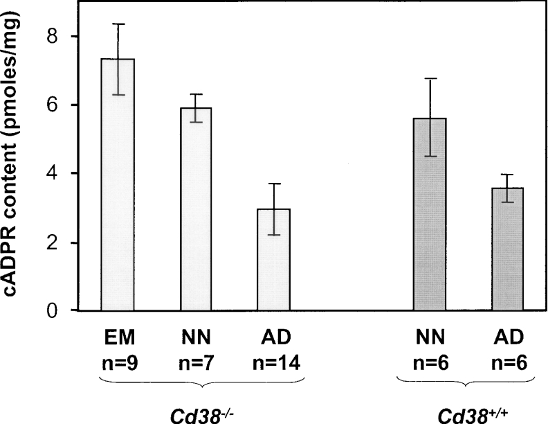
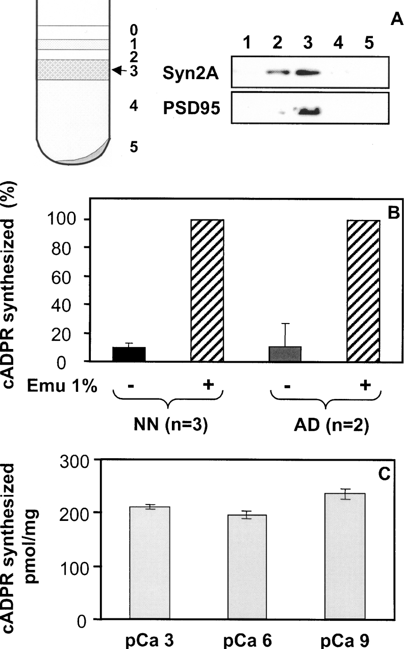
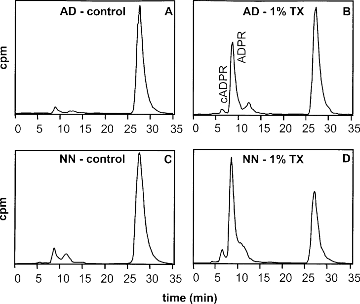
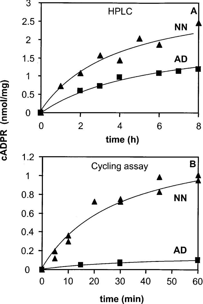
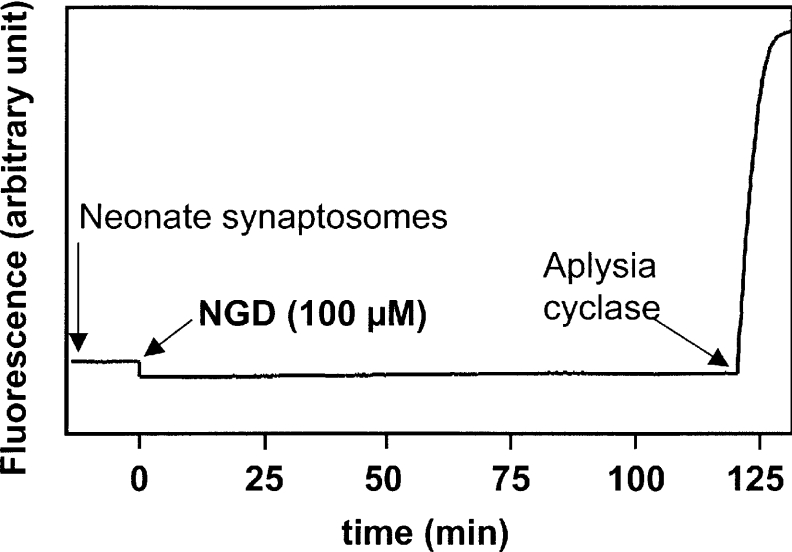
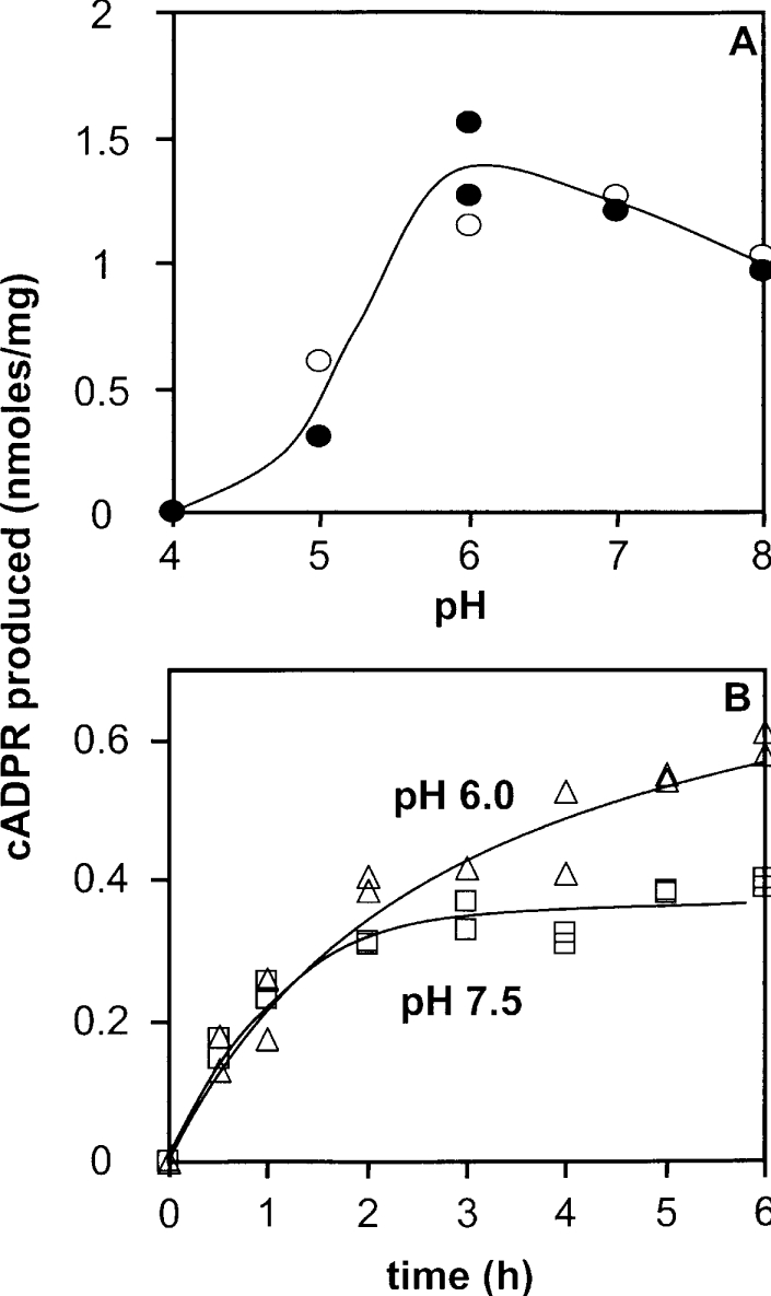
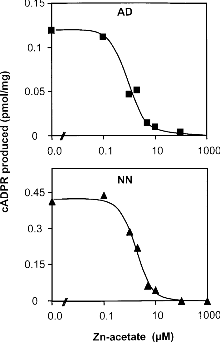
cADPR (cADP-ribose), a metabolite of NAD+, is known to modulate intracellular calcium levels and to be involved in calcium-dependent processes, including synaptic transmission, plasticity and neuronal excitability. However, the enzyme that is responsible for producing cADPR in the cytoplasm of neural cells, and particularly at the synaptic terminals of neurons, remains unknown. In the present study, we show that endogenous concentrations of cADPR are much higher in embryonic and neonate mouse brain compared with the adult tissue. We also demonstrate, by comparing wild-type and Cd38-/- tissues, that brain cADPR content is independent of the presence of CD38 (the best characterized mammalian ADP-ribosyl cyclase) not only in adult but also in developing tissues. We show that Cd38-/- synaptosome preparations contain high ADP-ribosyl cyclase activities, which are more important in neonates than in adults, in line with the levels of endogenous cyclic nucleotide. By using an HPLC method and adapting the cycling assay developed initially to study endogenous cADPR, we accurately examined the properties of the synaptosomal ADP-ribosyl cyclase. This intracellular enzyme has an estimated K(m) for NAD+ of 21 microM, a broad optimal pH at 6.0-7.0, and the concentration of free calcium has no major effect on its cADPR production. It binds NGD+ (nicotinamide-guanine dinucleotide), which inhibits its NAD+-metabolizing activities (K(i)=24 microM), despite its incapacity to cyclize this analogue. Interestingly, it is fully inhibited by low (micromolar) concentrations of zinc. We propose that this novel mammalian ADP-ribosyl cyclase regulates the production of cADPR and therefore calcium levels within brain synaptic terminals. In addition, this enzyme might be a potential target of neurotoxic Zn2+.


Figures

Figure 1
Figure 1. Endogenous cADPR content in developing and adult brain from wild-type and Cd38−/− mice
Extracts were prepared from tissues isolated from embryos (EM, days 15 or 16), from neonates (NN, days 1 or 2), or adults (AD), and were analysed for cADPR content using the cycling assay as described in the Experimental section. Results are means±S.D.
Figure 2
Figure 2. Analysis of the ADP-ribosyl cyclase activity from Cd38−/− mouse brain synaptosomes by the cycling assay: effects of detergent and free calcium concentration
(A) Synaptosomes preparation by centrifugation of brain homogenate on a discontinuous sucrose gradient and Western-blot analysis of the collected fractions containing proteins (6 μg/well) using anti-(synapsin IIa) and anti-PSD95 antibodies. Synaptosomes were collected at the 1.25 M/1.0 M sucrose interface (fraction 3; arrow). (B) Synaptosomes (0.4 mg/ml for adult and 0.2 mg/ml for neonate) were incubated (2 h for adult and 1 h for neonate) with 200 μM NAD+ in solution D (containing 10 mM Tris/HCl, pH 7.5, 2.2 mM CaCl2, 0.5 mM Na2HPO4, 0.4 mM KH2PO4, 4 mM NaHCO3 and 80 mM NaCl) in the absence (−) or presence (+) of the detergent Emulphogen (1%, w/v). Reactions were stopped by the addition of HClO4 followed by neutralization. The formation of cADPR was detected using the cycling assay as described in the Experimental section. The results obtained in the absence of detergent was expressed as percentages of the result obtained in the presence of the detergent. (C) Adult synaptosomes (0.4 mg/ml) were incubated for 2 h in 50 mM Tris/HCl, pH 7.5, in the presence of 200 μM NAD+ and 1% (w/v) Emulphogen, and with 1 nM (pCa 9), 1 μM (pCa 6) or 1 mM (pCa 3) free calcium. The experiment was reproduced twice with two different synaptosomal preparations (n=4). Results are means±S.D. Analysis of the cADPR production was performed using the cycling assay.
Figure 3
Figure 3. Analysis of NAD+-metabolizing activities of synaptosomes from adult and neonate Cd38−/− mice brain by HPLC: effects of detergent
Synaptosomes (1 mg/ml) prepared from brain tissue of adult (AD; A, B) and neonate (NN; C, D) Cd38−/− mice were incubated for 8 h in 20 mM Mes/NaOH at pH 6.0 and 32 °C with [14C]NAD+ (8 mCi/mmol) and NAD+ (final concentration 60 μM) in the presence of 4 mM EDTA, with (B, D) or without (A, C) 1% (w/v) Triton X-100 (TX). Reactions were stopped by the addition of HClO4 followed by neutralization. Products analysis was performed by HPLC using an online radioactivity detector as described in the Experimental section.
Figure 4
Figure 4. Kinetics of the ADP-ribosyl cyclase activity of synaptosomes from adult and neonate Cd38−/− brains, measured using both HPLC (A) and the cycling assay (B)
(A) Synaptosomes (1 mg of protein/ml) were incubated at 32 °C for the given times with [14C]NAD+ (8 mCi/mmol) and NAD+ (final concentration 60 μM), 4 mM EDTA, 1% (w/v) Triton X-100 in 20 mM Mes/NaOH at pH 6.0. (B) Synaptosomes [adult (AD) 0.4 mg/ml, neonate (NN) 0.2 mg/ml] were incubated for the given times with 200 μM NAD+ in solution D (see legend to Figure 3) in the presence of 1% (w/v) Emulphogen.
Figure 5
Figure 5. Properties of the CD38-independent ADP-ribosyl cyclase: determination of the Km (app) of NAD+ and of the Ki of NGD+
(A and B) Adult brain synaptosomes (0.4 mg/ml) were incubated for 15 or 30 min at 37 °C in solution D (pH 7.5) with 1% (w/v) Emulphogen and various NAD+ concentrations, in the absence (◇) or in the presence (×) of 100 μM NGD+. Then the cADPR formation was analysed using the cycling assay and reaction rates were estimated. (B) Secondary plot of the Km (app) for NAD+ against NGD+ concentration (n=3 at 0 NGD+; n=1 at 100 μM NGD+; n=2 at 300 μM NGD+). (C and D) Adult brain synaptosomes (1.1 mg of protein/ml) were incubated for 2 or 4 h at 37 °C in the presence of [14C]NAD+ (4 mCi/mmol) and NAD+ (60 μM), 4 mM EDTA, 1% (w/v) Triton X-100 in 20 mM Tris/HCl at pH 7.0 either in the absence or in the presence of 1 mM NGD+. Product analysis was performed by HPLC.
Figure 6
Figure 6. Absence of cGDPR formation by the ADP-ribosyl cyclase from Cd38−/− brain synaptosomes
NGD+ (100 μM) was added to 0.5 mg/ml neonate brain synaptosomes from Cd38−/− mice incubated at 37 °C in solution D. Formation of cGDPR was detected by fluorescence at 420 nm upon excitation at 300 nm. After 2 h of incubation, Aplysia cyclase (0.3 μg/ml) was added to fully transform the residual NGD+ into cGDPR (control).
Figure 7
Figure 7. pH effects on the CD38-independent synaptosomal ADP-ribosyl cyclase activity
(A) Adult brain synaptosomes (1 mg of protein/ml) were incubated for 6 h at 37 °C or 7 h at 32 °C in the presence of [14C]NAD+ (4 mCi/mmol) and NAD+ (60 μM), 4 mM EDTA, 1% (w/v) Triton X-100 in 20 or 40 mM citrate/HCl (pH 4.0), Mes/NaOH (pH 5.0 and 6.0), or Tris/HCl (pH 7.0 or 8.0). Product analysis was performed using HPLC. (B) Synaptosomes (adult, 0.4 mg/ml) were incubated for the given times with 60 μM NAD+ in solution D (see legend to Figure 3) containing either 30 mM Tris/HCl (pH 7.5) or 80 mM Mes/NaOH (pH 6.0) in the presence of 1% (w/v) Emulphogen. cADPR formation was analysed using the cycling assay.
Figure 8
Figure 8. Effect of Zn2+ on the ADP-ribosyl cyclase activity of synaptosomes from neonate and adult Cd38−/− mice
Synaptosomes (0.4 mg/ml for adult and 0.2 mg/ml for neonate) were incubated [2 h for adult (AD) and 1 h for neonate (NN)] with 200 μM NAD+ in solution D (see legend to Figure 2) containing 1% (w/v) Emulphogen and various concentrations of zinc acetate. The cADPR formation was analysed using the cycling assay.

Similar articles

Cited by

References

    1. Guse A. H. Cyclic ADP-ribose: a novel Ca2+-mobilising second messenger. Cell. Signalling. 1999;11:309–316. - PubMed
    1. Higashida H., Hashii M., Yokoyama S., Hoshi N., Chen X. L., Egorova A., Noda M., Zhang J. S. Cyclic ADP-ribose as a second messenger revisited from a new aspect of signal transduction from receptors to ADP-ribosyl cyclase. Pharmacol. Ther. 2001;90:283–296. - PubMed
    1. Rutter G. A. Calcium signalling: NAADP comes out of the shadows. Biochem. J. 2003;373:e3–e4. - PMC - PubMed
    1. Kinnear N. P., Boittin F. X., Thomas J. M., Galione A., Evans A. M. Lysosome–sarcoplasmic reticulum junctions: a trigger zone for calcium signaling by nicotinic acid adenine dinucleotide phosphate and endothelin-1. J. Biol. Chem. 2004;279:54319–54326. - PubMed
    1. Dammermann W., Guse A. H. Functional ryanodine receptor expression is required for NAADP-mediated local Ca2+ signaling in T-lymphocytes. J. Biol. Chem. 2005;280:21394–21399. - PubMed

Publication types

MeSH terms