

Involvement of 4-1BB (CD137)-4-1BBligand interaction in the modulation of CD4 T cell-mediated inflammatory colitis
- PMID: 16412046
- PMCID: PMC1809580
- DOI: 10.1111/j.1365-2249.2005.02991.x
Involvement of 4-1BB (CD137)-4-1BBligand interaction in the modulation of CD4 T cell-mediated inflammatory colitis
Abstract
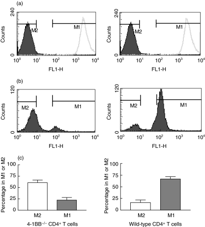
4-1BB ligand (4-1BBL) expressed on antigen-presenting cells interacts with 4-1BB on activated T cells (especially CD8+ cells) and co-stimulates the latter to secrete cytokines and to proliferate. The role of 4-1BB-4-1BBL interaction was studied here in a model of colitis based on naive CD4+ T cell transfer to SCID mice, a disease model in which CD8 cells do not take part. We found that CD4+ T cells from 4-1BB-deficient mice, after transfer in SCID mice, proliferated more rapidly compared to wild-type CD4+ T cells. Mice reconstituted with naive CD4+ T cells from 4-1BB-deficient mice developed colitis, however, with a mixed Th1/Th2 response, in contrast to the Th1-type response in mice reconstituted with wild-type naive CD4+ T cells. Importantly, this altered cytokine response did not temper colitis severity. Although it has been reported previously that 4-1BB co-stimulation may contribute to regulatory T cell functioning, we found that CD4+CD25+ regulatory T cells from 4-1BB-deficient mice were perfectly able to prevent naive CD4+ T cell-induced colitis. In conclusion, our data provide evidence that 4-1BB-4-1BBL interaction modulates the effector CD4+ T cell-driven immune response and cytokine production in experimental colitis without affecting regulatory T cell function.
Figures
References
-
- Vinay DS, Kwon BS. Role of 4-1BB in immune responses. Semin Immunol. 1998;10:481–9. - PubMed
-
- Alderson MR, Smith CA, Tough TW, et al. Molecular and biological characterization of human 4-1BB and its ligand. Eur J Immunol. 1994;24:2219–27. - PubMed
-
- Schwarz H, Valbracht J, Tuckwell J, von Kempis J, Lotz M. ILA, the human 4-1BB homologue, is inducible in lymphoid and other cell lineages. Blood. 1995;85:1043–52. - PubMed
-
- Kienzle G, von Kempis J. CD137 (ILA/4-1BB), expressed by primary human monocytes, induces monocyte activation and apoptosis of B lymphocytes. Int Immunol. 2000;12:73–82. - PubMed
-
- Melero I, Johnston JV, Shufford WW, Mittler RS, Chen L. NK1.1 cells express 4-1BB (CDw137) costimulatory molecule and are required for tumor immunity elicited by anti-4-1BB monoclonal antibodies. Cell Immunol. 1998;190:167–72. - PubMed
Publication types
MeSH terms
Substances
Grants and funding
LinkOut - more resources
Full Text Sources
Other Literature Sources
Molecular Biology Databases
Research Materials
