. 2006 Apr 10;347(2):410-21.
doi: 10.1016/j.virol.2005.11.044. Epub 2006 Jan 18.

Murine encephalitis caused by HCoV-OC43, a human coronavirus with broad species specificity, is partly immune-mediated

Affiliations

Murine encephalitis caused by HCoV-OC43, a human coronavirus with broad species specificity, is partly immune-mediated

Noah Butler et al. Virology. .

Abstract

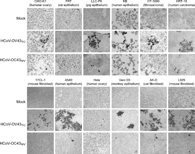
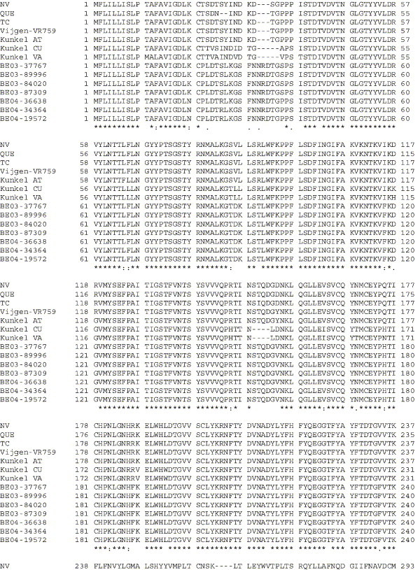
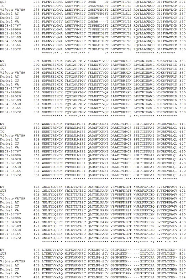
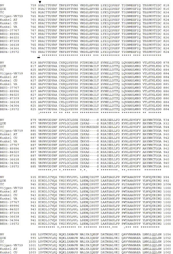
The human coronavirus HCoV-OC43 causes a significant fraction of upper respiratory tract infections. Most coronaviruses show a strong species specificity, although the SARS-Coronavirus crossed species from palm civet cats to infect humans. Similarly, HCoV-OC43, likely a member of the same coronavirus group as SARS-CoV, readily crossed the species barrier as evidenced by its rapid adaptation to the murine brain [McIntosh, K., Becker, W.B., Chanock, R.M., 1967. Growth in suckling-mouse brain of "IBV-like" viruses from patients with upper respiratory tract disease. Proc Natl Acad Sci U.S.A. 58, 2268-73]. Herein, we investigated two consequences of this plasticity in species tropism. First, we showed that HCoV-OC43 was able to infect cells from a large number of mammalian species. Second, we showed that virus that was passed exclusively in suckling mouse brains was highly virulent and caused a uniformly fatal encephalitis in adult mice. The surface glycoprotein is a major virulence factor in most coronavirus infections. We identified three changes in the HCoV-OC43 surface glycoprotein that correlated with enhanced neurovirulence in mice; these were located in the domain of the protein responsible for binding to host cells. These data suggest that some coronaviruses, including HCoV-OC43 and SARS-CoV, readily adapt to growth in cells from heterologous species. This adaptability has facilitated the isolation of HCoV-OC43 viral variants with markedly differing abilities to infect animals and tissue culture cells.


Figures

Fig. 1
Fig. 1
HCoV-OC43NV is uniformly lethal to wild type C57BL/6 mice. 5-week-old mice were inoculated intranasally with 30 LD50 HCoV-OC43NV or 106 TCID50 HCoV-OC43TC as described in Materials and methods. Mice were monitored daily for survival (A) and weight loss (B). (* Indicates P < 0.0002).
Fig. 2
Fig. 2
Viral titers and viral RNA burdens in the brains of HCoV-OC43NV-infected mice. Wild type and RAG1−/− C57BL/6 mice were infected intranasally with 30 LD50 HCoV-OC43NV. (A and B) Whole brains were isolated at the indicated time points and titers of infectious virus and virus RNA burden were determined as described in Materials and methods. Titers from 3 representative mice are shown for each time point demonstrating correlation between recovery of infectious virus and viral RNA burden. (C) HCoV-OC43 RNA titers increase until day 7 p.i. then decline by day 9. Whole RNA was isolated from the indicated tissue and assayed for virus burden with real-time RT-PCR. Data in panel C are expressed as mean ± SEM for 4–6 mice per time point. Note the different scales in “Brain” and “Spinal Cord” versus “Lung” and “Intestine.” (*Indicates P < 0.05).
Fig. 3
Fig. 3
Neurons are the primary target of HCoV-OC43NV in vivo. Mice were inoculated intranasally with 30 LD50 of HCoV-OC43NV and brains and spinal cords were harvested 7 or 9 days p.i. Tissue samples were prepared for in situ hybridization, antigen staining or Luxol Fast Blue (LFB) staining and slides were examined with standard light, fluorescence or confocal microscopy as described in Materials and methods. (A–B) HCoV-OC43 antigen is localized in the gray matter of spinal cords. Panel A depicts immunohistochemical detection of HCoV-OC43 using the anti-OC43 S hybridoma O.4.3. Panel B depicts a serial section stained with LFB to demarcate the white matter. (C–E) Morphology of HCoV-OC43NV-infected cells is consistent with that of neurons. Images are representative coronal sections of brain stems from mice harvested 7–9 days p.i. Sections were stained with O.4.3 followed by Cy3-labeled goat anti-mouse. (F–H) Combination in situ hybridization for HCoV-OC43 nucleocapsid RNA (Cy3-labeled antisense probe, red) and immunohistochemistry for the astrocyte marker GFAP (FITC-labeled anti-GFAP, green). Panel H is a merged image of panels F and G. Original images are 20× magnification for panels A and B, or 40× magnification for panels C–H.
Fig. 4
Fig. 4
T cell infiltration into the HCoV-OC43NV-infected CNS contributes to morbidity and mortality. (A) Both CD4 and CD8 T cells infiltrate the brains of HCoV-OC43NV-infected mice. Mononuclear cells were harvested 7 days p.i., surface stained for CD4 and CD8 and subjected to FACS analysis as described in Materials and methods. Data from one representative mouse are shown. (B) Two color FACS analysis of in vitro peptide-stimulated mononuclear cells demonstrating infiltration of OC43-specific CD4 T cells. Whole brains were isolated from HCoV-OC43NV-infected mice 7 days p.i. Mononuclear cells were surface stained for CD4 and intracellularly stained for IFN-γ ex vivo after no stimulation (left panel) or stimulation with M133 peptide (right panel). Data from one representative mouse are shown. (C) HCoV-OC43NV-infected RAG1−/− C57BL/6 mice lose weight (left panel) and succumb to infection (right panel) with delayed kinetics relative to infected wild type mice (*Indicates P < 0.05). (D) Relative burden of HCoV-OC43 RNA (left panel) and infectious virus (right panel) in brains of moribund wild type and RAG1−/− mice. Data in panel C are expressed as mean ± SEM for 4–6 mice per group. (*Indicates P < 0.05).
Fig. 5
Fig. 5
Broad tissue and cell type tropism of HCoV-OC43. A wide variety of tissue culture cells from hamster, rat, pig, human, mouse, monkey and cat were infected with 10 TCID50 (105 SMLD50) of HCoV-OC43NV, 30 TCID50 of HCoV-OC43TC or mock-infected as described in Materials and methods. After 3 days, virus-infected cells were detected with immunocytochemistry as described in Materials and methods. Nearly all of the cell lines were infected with both viruses, demonstrating that HCoV-OC43 exhibits a broad species specificity in vitro. Original images are 20× magnification.
Supplementary Fig. 1
Amino acid alignment of S protein sequences for HCoV-OC43NV, HCoV-OC43QUE, HCoV-OC43TC and the published sequence of several HCoV-OC43 isolates. Asterisks indicate identical residues, colons represent conserved changes and blank spaces denote non-conserved substitutions. The putative furin recognition site is underlined and the cleavage site is marked with an arrowhead. For HCoV-OC43NV and HCoV-OC43TC, total RNA was reverse transcribed to cDNA and the S gene was amplified by PCR. PCR products were directly sequenced as described in Materials and methods. The sequence for HCoV-OC43QUE corresponds to GenPept accession number AAT84362, passed 5–6 times in HRT-18 cells (St-Jean et al., 2004); the sequence for Vijgen VR-759 was translated from GenBank accession number AY391777, passed an unknown number of times in HRT-18 cells (Vijgen et al., 2005b); the sequence for Kunkel AT corresponds to GenPept accession number AAB27260, passed 3 times in HRT-18 cells (Kunkel and Herrler, 1993b); the sequences for Kunkel CU and Kunkel VA correspond to GenPept accession numbers CAA83660 and CAA83661, passed 3 times in MDCK cells and 12 times in Vero E6 cells, respectively (Kunkel and Herrler, 1996); and sequences for HCoV-OC43 clinical isolates BE03-37767, BE03-89996, BE03-84020, BE03-87309, BE04-36638, BE04-34364 and BE04-19572 correspond to GenPept accession numbers AAX84794, AAX84791, AAX84793, AAX85674, AAX84795, AAX84792 and AAX85678, respectively, submitted by Vijgen et al. (2005a).

References

    1. Akashi H., Inaba Y., Miura Y., Sato K., Tokuhisa S., Asagi M., Hayashi Y. Propagation of the Kakegawa strain of bovine coronavirus in suckling mice, rats and hamsters. Arch. Virol. 1981;67(4):367–370. - PMC - PubMed
    1. Barthold S.W., de Souza M.S., Smith A.L. Susceptibility of laboratory mice to intranasal and contact infection with coronaviruses of other species. Lab. Anim. Sci. 1990;40(5):481–485. - PubMed
    1. Bergmann C.C., Altman J.D., Hinton D., Stohlman S.A. Inverted immunodominance and impaired cytolytic function of CD8+T cells during viral persistence in the central nervous system. J. Immunol. 1999;163:3379–3387. - PubMed
    1. Binder G.K., Griffin D.E. Interferon-gamma-mediated site-specific clearance of alphavirus from CNS neurons. Science. 2001;293(5528):303–306. - PubMed
    1. Byrnes A.P., Griffin D.E. Binding of Sindbis virus to cell surface heparan sulfate. J. Virol. 1998;72(9):7349–7356. - PMC - PubMed

LinkOut - more resources