

doi: 10.1128/JVI.80.3.1152-1159.2006.
Transduction of nondividing human macrophages with gammaretrovirus-derived vectors
Affiliations
- PMID: 16414992
- PMCID: PMC1346929
- DOI: 10.1128/JVI.80.3.1152-1159.2006
Item in Clipboard
Transduction of nondividing human macrophages with gammaretrovirus-derived vectors
J Virol.
2006 Feb.
Abstract
It is commonly accepted that infection of nondividing cells by gammaretroviruses such as the murine leukemia viruses is inefficient due to their inability to cross the nuclear envelope barrier. Challenging this notion, we now show that human nondividing macrophages display a specific window of susceptibility to transduction with a Friend murine leukemia virus (F-MLV)-derived vector during their differentiation from monocytes. This finding suggests that factors other than the nuclear membrane govern permissiveness to gammaretroviral infection and raises the possibility of using the macrophage tropism of F-MLV in gene therapy.
Figures

Schematic representation of the retroviral vectors used here. The HIV-1 (22, 28) and F-MLV (42) vectors are shown by open and gray boxes, respectively. For simplicity, only the viral accessory proteins coded for by the HIV-1 packaging construct (all except Vpu) are drawn in the figure; the main viral elements of each transfer vector are shown as follows: ψ, packaging sequence; RRE, Rev-responsive element; cPPT, central polypurine tract; U3*, partially deleted and self-inactivating LTR; SD and SA, slice donor and acceptor sites, respectively. Both vectors express GFP driven from a CMV promoter.
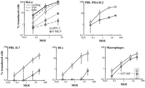
Transduction of growth-arrested HeLa cells and of primary human blood cells with HIV-1 and F-MLV vectors. VSVg-pseudotyped vectors were purified on a double-sucrose cushion after production from 293T cells, and normalized amounts of HeLa infectious particles were used on target cells at different MOIs (as indicated). The percentage of GFP-positive cells was scored 3 to 4 days later by flow cytometry. The different primary cells shown here were derived from the same donor, and average results from three to seven different donors are presented. HeLa cells were arrested in either G2/M by gamma irradiation (γ-irr; 6,000 rads) or in G1/S by aphidicolin (aphi) treatment (10 μg/ml), and PBLs were treated with PHA plus IL-2 (to induce cell proliferation) or IL-7 (to induce G0 to G1b transition) 24 h prior to transduction. Human blood monocytes were differentiated in macrophages and DCs in presence of GM-CSF and GM-CSF plus IL-4, respectively, and transduced between days 4 and 6. When indicated, AZT and ddI were added to cells prior to viral transduction.
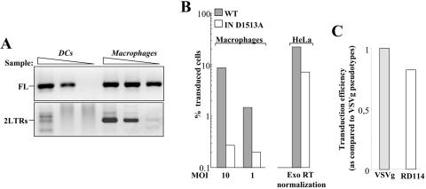
Proviral DNA analysis and requirement for IN activity in the transduction of human macrophages. (A) Day 4 DCs and macrophages were transduced with the same amount of F-MLV vectors at an MOI 10 and lysed 24 h postinfection for the analysis of late reverse transcription products that were full-length (FL) and 2LTR circles. Analysis was performed on threefold dilution of the sample with primers that allow the specific amplification of each form followed by agarose gel migration and detection. (B) A single point mutation was introduced in the catalytic aspartic acid of the F-MLV IN (D1513A) by site-directed mutagenesis. Mutant and wild-type (WT) viruses were produced in parallel, and their infectivity was tested on growing HeLa cells after normalization by exogenous (Exo) RT activity (right panel). Upon normalization of their infectious titer, wild-type and mutant viral preparations were used to infect macrophages (left panel). (C) VSVg- and RD114-pseudotyped MLV vectors were similarly produced and tested on macrophages at equal MOIs. The infectivity of RD114-pseudotyped MLV vectors is shown with respect to that of VSVg-MLV, set arbitrarily at 1.

Features of F-MLV-transduced macrophages. (A) Transmission and fluorescence microscopy pictures of plated F-MLV-transduced macrophage cultures. (B) Cell surface marker analysis 2 days posttransduction of day 4 macrophages. Macrophages were incubated with the indicated fluorescently coupled antibody prior to flow cytometry analysis. (C) To demonstrate that viral transduction occurred in differentiated macrophages, day 4 macrophages were incubated with PE-labeled latex beads for 2 h followed by extensive cell washing to remove unbound beads. Two hours afterwards, the cell culture was infected. Cells were analyzed by fluorescence microscopy 3 days postinfection.

Comparison between Moloney MLV and Friend MLV-derived retroviral vectors. The Friend MLV-derived vector was compared to a Moloney MLV-derived vector produced in parallel. This vector has been previously described elsewhere (23, 24), and it is comparable to the Friend MLV-derived vector used here. After normalization of their infectious titers on HeLa cells, equal MOIs were used in the transduction of day 4 macrophages, and infection was scored 3 days later by flow cytometry analysis.

Absence of cell proliferation in human GM-CSF-derived macrophages. (A) Monocytes (gamma irradiated [γ-irr.] as a control or not, both with GM-CSF) and cycling Jurkat T cells were seeded in equal numbers in the presence of [3H]thymidine for 15 days prior to analysis. (B) Monocytes and PBLs were labeled with CFSE at day 0 and then differentiated with GM-CSF or stimulated with CD3 or CD28 plus IL-2 for 8 days, respectively, prior to flow cytometry analysis. The graph shows a comparison between CFSE fluorescence at day 0 and that at day 8, as indicated. (C) Day 4 GM-CSF-derived macrophages and Jurkat T cells were incubated for 24 h in BrdU, prior to staining with a fluorescently labeled anti-BrdU antibody and flow cytometry analysis. In each histogram plot, BrdU-negative and BrdU-positive samples (continuous and dotted lines, respectively) are superimposed.

Susceptibility of macrophages to retroviral transduction is time and culture condition dependent. (A) Monocytes were differentiated into macrophages in GM-CSF, M-CSF, or human serum (hAB) prior to viral infection at an MOI of 25 and flow cytometry analysis. (B) Time curve susceptibility to retroviral transduction of GM-CSF-derived macrophages. Time zero indicates cells immediately after the 3-h serum deprivation step and addition of GM-CSF and FCS. The results presented here represent the average of three independent experiments with cells of different donors.
References
-
- Burke, B. 2003. Macrophages as novel cellular vehicles for gene therapy. Expert Opin. Biol. Ther. 3:919-924. - PubMed
-
- Dardalhon, V., S. Jaleco, S. Kinet, B. Herpers, M. Steinberg, C. Ferrand, D. Froger, C. Leveau, P. Tiberghien, P. Charneau, N. Noraz, and N. Taylor. 2001. IL-7 differentially regulates cell cycle progression and HIV-1-based vector infection in neonatal and adult CD4+ T cells. Proc. Natl. Acad. Sci. USA 98:9277-9282. - PMC - PubMed
-
- de Noronha, C. M., M. P. Sherman, H. W. Lin, M. V. Cavrois, R. D. Moir, R. D. Goldman, and W. C. Greene. 2001. Dynamic disruptions in nuclear envelope architecture and integrity induced by HIV-1 Vpr. Science 294:1105-1108. - PubMed
Publication types
MeSH terms
Substances
LinkOut - more resources
Full Text Sources
Other Literature Sources
