. 2006 Feb;80(3):1290-301.
doi: 10.1128/JVI.80.3.1290-1301.2006.

West Nile virus discriminates between DC-SIGN and DC-SIGNR for cellular attachment and infection

Affiliations

West Nile virus discriminates between DC-SIGN and DC-SIGNR for cellular attachment and infection

Carl W Davis et al. J Virol. 2006 Feb.

Abstract

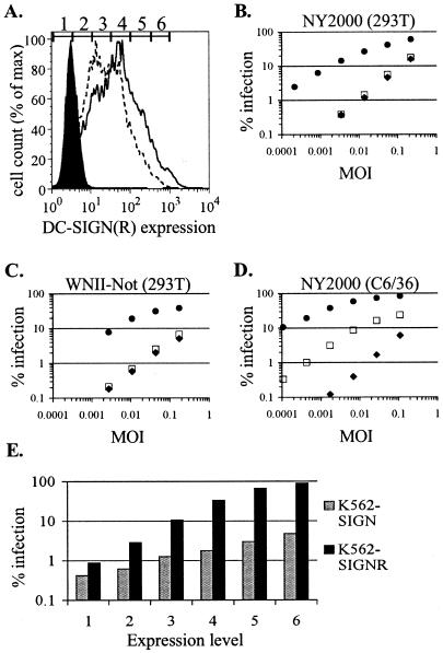
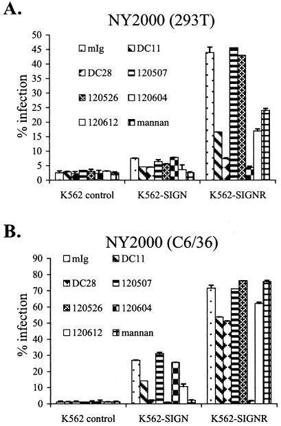
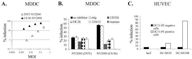
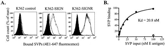
The C-type lectins DC-SIGN and DC-SIGNR bind mannose-rich glycans with high affinity. In vitro, cells expressing these attachment factors efficiently capture, and are infected by, a diverse array of appropriately glycosylated pathogens, including dengue virus. In this study, we investigated whether these lectins could enhance cellular infection by West Nile virus (WNV), a mosquito-borne flavivirus related to dengue virus. We discovered that DC-SIGNR promoted WNV infection much more efficiently than did DC-SIGN, particularly when the virus was grown in human cell types. The presence of a single N-linked glycosylation site on either the prM or E glycoprotein of WNV was sufficient to allow DC-SIGNR-mediated infection, demonstrating that uncleaved prM protein present on a flavivirus virion can influence viral tropism under certain circumstances. Preferential utilization of DC-SIGNR was a specific property conferred by the WNV envelope glycoproteins. Chimeras between DC-SIGN and DC-SIGNR demonstrated that the ability of DC-SIGNR to promote WNV infection maps to its carbohydrate recognition domain. WNV virions and subviral particles bound to DC-SIGNR with much greater affinity than DC-SIGN. We believe this is the first report of a pathogen interacting more efficiently with DC-SIGNR than with DC-SIGN. Our results should lead to the discovery of new mechanisms by which these well-studied lectins discriminate among ligands.


Figures

FIG. 1.
FIG. 1.
WNV preferentially infects K562 cell lines expressing DC-SIGNR. (A) Histogram analysis of DC-SIGN(R) expression in K562 cell lines. Cells were stained with DC11-PE and analyzed by flow cytometry. Filled area, control cells; dotted line, K562-SIGN cells; solid line, K562-SIGNR cells. Numbers and gates shown above histograms define the expression levels referred to in panel E. (B) K562-SIGN (open squares), K562-SIGNR (closed circles), or control K562 (closed diamonds) cells were infected with serial fourfold dilutions of lineage I WNV strain NY2000 grown in HEK 293T cells. Percentages of infection were assessed 20 h later by intracellular FACS staining for WNV E protein. Similar results were observed in more than five separate experiments. (C) Infection of K562 cells was performed as described for panel B, except cells were infected with lineage II WNV strain WNII-Not grown in HEK 293T cells. This strain lacks an N-linked glycosylation site on the viral E protein. A representative experiment of three performed is shown. (D) Infection was performed as described for panel B, except cells were infected with NY2000 grown in C6/36 mosquito cells. (E) K562-SIGN and K562-SIGNR cells were infected with 293T-derived virus at an MOI of 0.004. DC11-PE was included during staining for E antigen to assess DC-SIGN(R) expression. Cells were grouped by expression level according to the gates defined in panel A, and infection was assessed in each subset.
FIG. 2.
FIG. 2.
Specific inhibitors prevent DC-SIGN(R)-mediated enhancement of WNV infection. (A) K562 cell lines were incubated with the indicated inhibitors for 30 min at 37°C and infected with 293T-derived NY2000 at an MOI of 0.02. Inhibitors were included in the virus input to keep the concentration of inhibitor constant. Infections were performed without removal of viral input or inhibitors, and percentages of infection were assessed by intracellular FACS after 20 h. Antibodies were used at a concentration of 5 μg/ml and mannan was used at 500 μg/ml. mIg refers to control mouse serum IgG. MAbs DC11 and DC28 are DC-SIGN(R) cross-reactive and bind to the repeat domain, 120526 and 120612 are DC-SIGN(R) cross-reactive and bind to the CRD, 120507 binds to the CRD of DC-SIGN only, and 120604 binds to the CRD of DC-SIGNR only. Values shown are the averages from duplicate wells with standard deviations indicated. A representative experiment of two performed is shown. (B) Infections were performed as described for panel A, except that C6/36-derived NY2000 was used.
FIG. 3.
FIG. 3.
DC-SIGN(R)-mediated enhancement of infection in primary cell cultures. (A) Titration of 293T-derived (open triangles) and C6/36-derived (closed triangles) WNV strain NY2000 on immature MDDCs. Percentages of infection were assessed after 20 h by intracellular FACS. Similar results were obtained using MDDCs from two other donors. (B) Blockade of MDDC infection by inhibitors of DC-SIGN. MDDCs were preincubated with the indicated inhibitors and infected with 293T- or C6/36-derived NY2000. Percentages of infection were assessed after 20 h by intracellular FACS. MAbs were used at 10 μg/ml, and mannan was used at 500 μg/ml. Infections were performed at an MOI of 0.09 for NY2000 grown in 293T cells and at an MOI of 0.04 for C6/36-derived virus. One representative experiment out of two performed is shown. (C) HUVECs were infected with lentiviruses encoding a control gene (lacZ), DC-SIGN, or DC-SIGNR at an MOI of 0.1. Seventy-two h posttransduction, cells were infected with 293T-derived NY2000 at an MOI of 0.05. After 20 h, intracellular staining for WNV E protein and DC-SIGN(R) expression was performed. For HUVECs transduced with DC-SIGN or DC-SIGNR lentiviruses, percentages of infection were assessed in both DC-SIGN(R)-expressing (DC11-PE-positive) and nonexpressing (DC11-PE-negative) cell populations.
FIG. 4.
FIG. 4.
WNV SVPs produced in human cells and containing native glycosylation patterns bind selectively to cells expressing DC-SIGNR. (A) Binding of WNV SVPs to K562 cells. The indicated cell lines were incubated with medium only (filled area) or with NY99-6480 SVPs (6 nM E protein). SVPs were produced in 293T cells under standard conditions (solid lines) to produce particles with native glycosylation patterns or in the presence of the Golgi mannosidase inhibitor 1-deoxymannojirimycin (dashed lines) to increase the incorporation of high-mannose N-linked glycans. Bound SVPs were detected by staining with anti-WNV MAb 4E1-647. (B) K562-SIGNR cells (circles) were incubated with serial twofold dilutions of SVPs produced under standard conditions and processed for FACS as described for panel A. The geometric mean 4E1-647 fluorescence was calculated at each concentration of SVPs, and the background fluorescence of cells incubated in the absence of SVPs was subtracted from each value. A one-site binding curve was fit to these data and is shown as a solid line, with the dissociation constant (Kd) indicated. For reference, the background-subtracted binding to K562 control cells (diamond) is shown at the highest SVP input.
FIG. 5.
FIG. 5.
The selective usage of DC-SIGNR for infection is specific to WNV glycoproteins containing native glycosylation patterns. A Renilla luciferase-expressing WNV replicon was packaged into RVPs by transfection of BHK cells containing this replicon with expression plasmids encoding flavivirus capsid proteins and prM-E polyproteins. Serial fourfold dilutions of RVPs were added to K562 cell lines, and infection was assessed after 48 h by measuring luciferase activity. Open squares, K562-SIGN cells; closed circles, K562-SIGNR cells; closed diamonds, K562 control cells. (A) RVPs were made by use of pWNIIcap, encoding the WNV capsid protein, and pCBWN, encoding the prM-E polyprotein of WNV strain NY99-6480 (NY99). The E protein of NY99-6480 is identical to the E protein of NY2000. (B) RVPs were made using pDEN1cap, encoding the dengue virus capsid protein, and pDV1 prM-E VAX, encoding a serotype 1 dengue virus prM-E polyprotein. (C) RVPs were made as described for panel A, but DMJ was added during production to yield particles containing predominantly high-mannose N-linked glycans. Similar results were seen with more than four separate RVP preparations. Note the difference between the x axis scale in panels A and C and that in panel B.
FIG. 6.
FIG. 6.
The use of DC-SIGNR as an attachment factor by WNV requires at least one N-linked glycan on either prM or E. RVPs were produced by transfection of replicon-containing BHK cells with pWNIICap and different WNV prM-E expression plasmids (as indicated above the graphs) and used to infect K562 cell lines. Infection was assessed after 48 h by measuring luciferase activity. Open squares, K562-SIGN cells; closed circles, K562-SIGNR cells; closed diamonds, K562 control cells. prM-E and capsid expression plasmids were transfected alone (A through D) or cotransfected with an expression plasmid encoding human furin (E through H). “NY99 prM KO” refers to pCBWN containing an asparagine-to-glutamine substitution at prM residue 15 which resulted in the removal of the N-linked glycosylation site. “NY99 E KO” contains an asparagine-to-glutamine substitution removing the N-linked site at E protein residue 154. “NY99 prM/E KO” contains both prM N15Q and E N154Q substitutions. “WNII-Not” here refers to plasmid pWNIIprM-E, containing the prM-E polyprotein from lineage II WNV strain WNII-Not. This strain lacks an N-linked glycosylation site on the E protein but still possesses the site on prM. A representative experiment out of two performed for each RVP preparation is shown. Similar results were seen with two separate RVP preparations. Note the difference between the x axis scale in panels A and E and that in the other panels.
FIG. 7.
FIG. 7.
Specific utilization of DC-SIGNR by WNV requires the DC-SIGNR CRD. (A) Schematic of DC-SIGN(R) domain structure. C, cytoplasmic domain (amino acids 1 to 41 of DC-SIGN/amino acids 1 to 49 of DC-SIGNR); TM, transmembrane domain (42 to 60/50 to 72); N, N-terminal domain (61 to 80/73 to 92); NLG, N-linked glycosylation site at asparagine (80/92); Repeat domain (81 to 252/93 to 264); CRD (253 to 404/265 to 399). (B) Expression of DC-SIGN(R) chimeras in stable K562 cell lines. Chimeras are named according to their five-domain compositions by the domain classifications given above. For example, chimera RRRSS consists of the DC-SIGNR cytoplasmic, transmembrane, and N-terminal domains fused to the repeat domain and the CRD of DC-SIGN. Cells were stained with DC11-PE and analyzed by flow cytometry. The geometric mean DC11-PE fluorescence is shown. (C) K562 cell lines expressing the indicated chimeras were infected with serial fourfold dilutions of 293T-derived NY2000. Percentages of infection were assessed by intracellular FACS after 20 h.

References

    1. Albelda, S. M., M. Daise, E. M. Levine, and C. A. Buck. 1989. Identification and characterization of cell-substratum adhesion receptors on cultured human endothelial cells. J. Clin. Investig. 83:1992-2002. - PMC - PubMed
    1. Allison, S. L., K. Stadler, C. W. Mandl, C. Kunz, and F. X. Heinz. 1995. Synthesis and secretion of recombinant tick-borne encephalitis virus protein E in soluble and particulate form. J. Virol. 69:5816-5820. - PMC - PubMed
    1. Alvarez, C. P., F. Lasala, J. Carrillo, O. Muniz, A. L. Corbi, and R. Delgado. 2002. C-type lectins DC-SIGN and L-SIGN mediate cellular entry by Ebola virus in cis and in trans. J. Virol. 76:6841-6844. - PMC - PubMed
    1. Baribaud, F., R. W. Doms, and S. Pohlmann. 2002. The role of DC-SIGN and DC-SIGNR in HIV and Ebola virus infection: can potential therapeutics block virus transmission and dissemination? Expert Opin. Ther. Targets 6:423-431. - PubMed
    1. Baribaud, F., S. Pohlmann, G. Leslie, F. Mortari, and R. W. Doms. 2002. Quantitative expression and virus transmission analysis of DC-SIGN on monocyte-derived dendritic cells. J. Virol. 76:9135-9142. - PMC - PubMed

Publication types

MeSH terms

LinkOut - more resources