. 2006 Feb;80(3):1440-50.
doi: 10.1128/JVI.80.3.1440-1450.2006.

Role of helix P of the human cytomegalovirus DNA polymerase in resistance and hypersusceptibility to the antiviral drug foscarnet

Affiliations

Role of helix P of the human cytomegalovirus DNA polymerase in resistance and hypersusceptibility to the antiviral drug foscarnet

Egor P Tchesnokov et al. J Virol. 2006 Feb.

Abstract

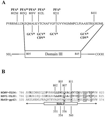
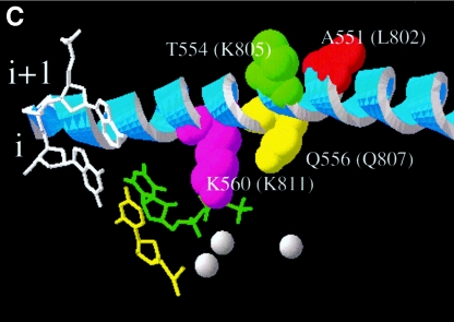
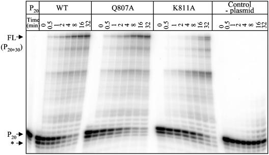
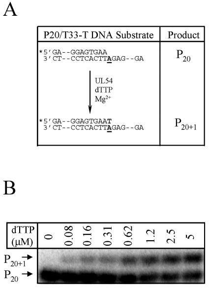
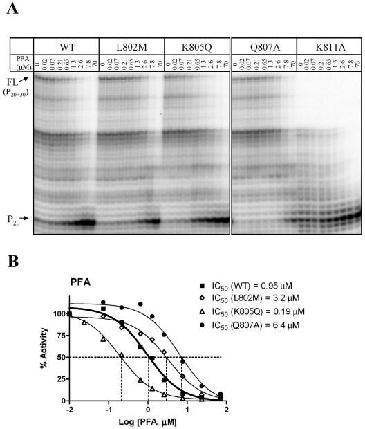
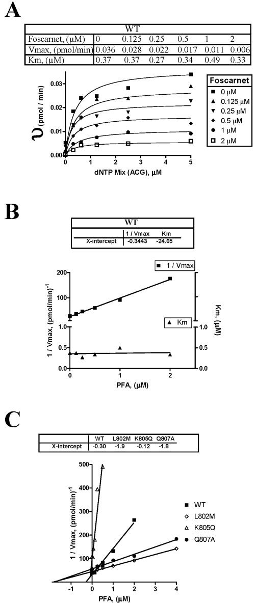
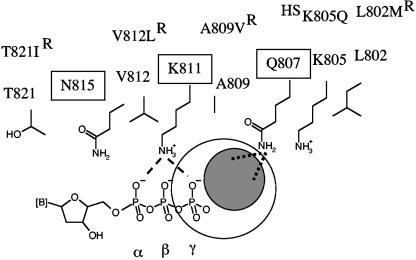
Mutations in the human cytomegalovirus DNA polymerase (UL54) can not only decrease but also increase susceptibility to the pyrophosphate (PP(i)) analogue foscarnet. The proximity of L802M, which confers resistance, and K805Q, which confers hypersusceptibility, suggests a possible unifying mechanism that affects drug susceptibility in one direction or the other. We found that the polymerase activities of L802M- and K805Q-containing mutant enzymes were literally indistinguishable from that of wild-type UL54; however, susceptibility to foscarnet was decreased or increased, respectively. A comparison with the crystal structure model of the related RB69 polymerase suggests that L802 and K805 are located in the conserved alpha-helix P that is implicated in nucleotide binding. Although L802 and K805 do not appear to make direct contacts with the incoming nucleotide, it is conceivable that changes at these residues could exert their effects through the adjacent, highly conserved amino acids Q807 and/or K811. Our data show that a K811A substitution in UL54 causes reductions in rates of nucleotide incorporation. The activity of the Q807A mutant is only marginally affected, while this enzyme shows relatively high levels of resistance to foscarnet. Based on these data, we suggest that L802M exerts its effects through subtle structural changes in alpha-helix P that affect the precise positioning of Q807 and, in turn, its presumptive involvement in binding of foscarnet. In contrast, the removal of a positive charge associated with the K805Q change may facilitate access or increase affinity to the adjacent Q807.


Figures

FIG.1.
FIG.1.
Location of resistance-conferring mutations in helix P. (A) Amino acid sequence of conserved domain III of the HCMV DNA polymerase UL54. Highlighted are residues that have been associated with changes in drug susceptibility: cidofovir (CDV), ganciclovir (GCV), and foscarnet (PFA). Superscript R and HS indicate resistant and hypersusceptible phenotypes, respectively. (B) Amino acid sequence of helix P of the RB69-associated DNA polymerase gp43 aligned against corresponding sequences of UL54 (HCMV) and UL30 (HSV1). Bold letters indicate amino acid residues that are conserved in both UL54 and UL30. Underlined letters indicate amino acid residues conserved among all three related DNA polymerases. Highly conserved residues are found between positions 807 and 822. (C) Interaction between helix P and the bound nucleotide, based on the structure of the ternary complex of gp43 (14). Divalent metal ions are shown as gray spheres. The bound nucleotide is shown in green, the position of the 3′ end of the primer is shown in yellow, and the complementary template positions i + 1 and i are shown in white. Helix P (A535 to G571) is shown in blue, and residues that are relevant to this work are highlighted: A551 (red; changes at this position in UL54 are associated with resistance to foscarnet), T554 (green; changes at this position in UL54 are associated with hypersusceptibility to foscarnet), Q556 (yellow), and K560 (magenta). The last two amino acids are highly conserved among α-DNA polymerases. Amino acids in parentheses indicate the structural equivalents in UL54.
FIG.1.
FIG.1.
Location of resistance-conferring mutations in helix P. (A) Amino acid sequence of conserved domain III of the HCMV DNA polymerase UL54. Highlighted are residues that have been associated with changes in drug susceptibility: cidofovir (CDV), ganciclovir (GCV), and foscarnet (PFA). Superscript R and HS indicate resistant and hypersusceptible phenotypes, respectively. (B) Amino acid sequence of helix P of the RB69-associated DNA polymerase gp43 aligned against corresponding sequences of UL54 (HCMV) and UL30 (HSV1). Bold letters indicate amino acid residues that are conserved in both UL54 and UL30. Underlined letters indicate amino acid residues conserved among all three related DNA polymerases. Highly conserved residues are found between positions 807 and 822. (C) Interaction between helix P and the bound nucleotide, based on the structure of the ternary complex of gp43 (14). Divalent metal ions are shown as gray spheres. The bound nucleotide is shown in green, the position of the 3′ end of the primer is shown in yellow, and the complementary template positions i + 1 and i are shown in white. Helix P (A535 to G571) is shown in blue, and residues that are relevant to this work are highlighted: A551 (red; changes at this position in UL54 are associated with resistance to foscarnet), T554 (green; changes at this position in UL54 are associated with hypersusceptibility to foscarnet), Q556 (yellow), and K560 (magenta). The last two amino acids are highly conserved among α-DNA polymerases. Amino acids in parentheses indicate the structural equivalents in UL54.
FIG. 2.
FIG. 2.
Efficiency of DNA synthesis with WT and mutant enzymes. The levels of DNA synthesis were measured with the heteropolymeric primer/template substrate P20/T50 DNA as a function of time. FL denotes the full-length product, and P20 denotes the unextended primer. The asterisk points to the exonuclease activity of UL54 enzymes. This band is less pronounced in the control without enzyme (left, primer only) and in reaction mixtures that contain plasmid pCITE4b without the coding sequence for UL54 (right).
FIG. 3.
FIG. 3.
Steady-state kinetic analyses. (A) Graphic representation of the experimental setup used to monitor single-nucleotide incorporations. The substrate P20/T33-T DNA allows the incorporation of dTTP, which extends primer P20 by a single nucleotide to yield P20 + 1. Complete sequences are shown in Materials and Methods. (B) Incorporation of dTMP by WT UL54. efficiency of single-nucleotide incorporations was monitored in the presence of increasing concentrations of dTTP, while all other deoxyribonucleotides were omitted from the reaction. The reaction was allowed to proceed for 10 min. Under these conditions, no significant extensions in the control reactions that contain plasmid pCITE4b without the coding sequence for UL54 were seen. This approach was used to determine the kinetic parameters that are listed in Table 1.
FIG. 4.
FIG. 4.
Measurements of IC50 values for inhibition of DNA synthesis in the presence of foscarnet. (A) The amount of full-length (FL) DNA synthesis was measured in the presence of increasing concentrations of foscarnet as indicated. Reactions were allowed to proceed for 30 min. (B) Dose-response curves and measurements of IC50 values for inhibition of DNA synthesis by WT and mutant enzymes. Changes in the ratios of the full-length product and the unextended primer were quantified to generate dose-response curves. Dotted lines point to the concentration of the inhibitor that reduces the activity of the enzyme by 50% (IC50).
FIG. 5.
FIG. 5.
Type of inhibition and measurements of Ki values. (A) DNA synthesis was measured at a single time point (30 min) in the presence of increasing concentrations of each the four nucleotides. This experiment was repeated with different concentrations of foscarnet to determine Ki values as described in Materials and Methods. This figure shows changes in Vmax and Km for WT UL54 as an example. υ, velocity of the reaction. The data for WT and mutant enzymes are summarized in Table 3. (B) Replot of 1/Vmax and Km values determined as for panel A, shown here as a function of foscarnet concentration. The x intercept provides the Ki value. Changes in Vmax at constant Km point to a noncompetitive type of inhibition. This is shown for WT UL54 as an example. (C) Values of 1/Vmax determined as for panel A and replotted as for panel B, shown here for WT and mutant enzymes.
FIG. 6.
FIG. 6.
Possible interactions between helix P and foscarnet. This simplified model shows side chains of important residues in helix P that are either highly conserved (boxed) among α-DNA polymerases or associated with changes in susceptibility to the drug (R, resistant; HS, hypersusceptible). Helix P of UL54 is shown here in an orientation to the bound nucleotide similar to that shown for helix P of gp43 in Fig. 1C. The structural data suggest that positions 805 and 802 are likely to be too far away to provide direct contacts with the drug. Our data suggest that foscarnet may bind in close proximity to Q807. The gray circle indicates the possible location of the bound drug relative to the nucleotide. The orientation of the bound foscarnet cannot be predicted on the basis of the data presented in this study. Dotted lines illustrate possible interactions between the negatively charged foscarnet and the side chain of Q807. There might be a partial overlap with the position of the γ-phosphate (large open circle), which would help to explain previous data that suggested a competitive mode of inhibition of pyrophosphorolysis. Dashed lines illustrate contacts between the structural equivalent of K811 (K560) in gp43 and the α- and γ-phosphates of the bound dNTP. Possible interaction between K811 and foscarnet cannot be confirmed, because mutations at this position were shown to affect dNTP binding at the same time.

References

    1. Bestman-Smith, J., and G. Boivin. 2003. Drug resistance patterns of recombinant herpes simplex virus DNA polymerase mutants generated with a set of overlapping cosmids and plasmids. J. Virol. 77:7820-7829. - PMC - PubMed
    1. Boosalis, M. S., J. Petruska, and M. F. Goodman. 1987. DNA polymerase insertion fidelity. Gel assay for site-specific kinetics. J. Biol. Chem. 262:14689-14696. - PubMed
    1. Chou, S., N. S. Lurain, K. D. Thompson, R. C. Miner, and W. L. Drew. 2003. Viral DNA polymerase mutations associated with drug resistance in human cytomegalovirus. J. Infect. Dis. 188:32-39. - PubMed
    1. Chou, S. W. 2001. Cytomegalovirus drug resistance and clinical implications. Transpl. Infect. Dis. 3(Suppl. 2):20-24. - PubMed
    1. Cihlar, T., M. D. Fuller, and J. M. Cherrington. 1998. Characterization of drug resistance-associated mutations in the human cytomegalovirus DNA polymerase gene by using recombinant mutant viruses generated from overlapping DNA fragments. J. Virol. 72:5927-5936. - PMC - PubMed

Publication types

MeSH terms

LinkOut - more resources