Comparative Study
. 2006 Feb;80(3):1463-75.
doi: 10.1128/JVI.80.3.1463-1475.2006.

Infectious molecular clones from a simian immunodeficiency virus-infected rapid-progressor (RP) macaque: evidence of differential selection of RP-specific envelope mutations in vitro and in vivo

Affiliations
Comparative Study

Infectious molecular clones from a simian immunodeficiency virus-infected rapid-progressor (RP) macaque: evidence of differential selection of RP-specific envelope mutations in vitro and in vivo

Takeo Kuwata et al. J Virol. 2006 Feb.

Abstract

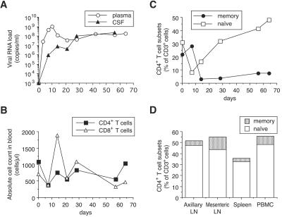
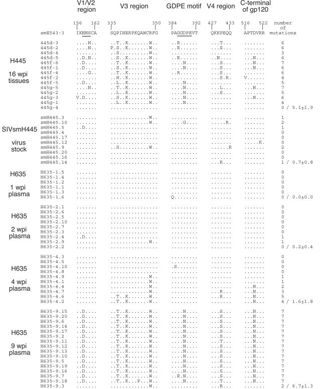
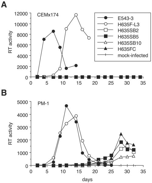
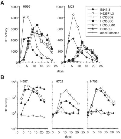
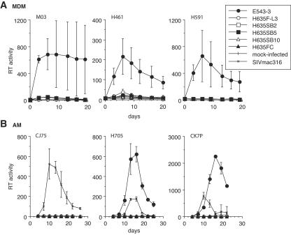
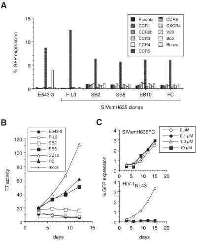
A minor fraction of simian immunodeficiency virus (SIV)-infected macaques progress rapidly to AIDS in the absence of SIV-specific immune responses. Common mutations in conserved residues of env in three SIVsmE543-3-infected rapid-progressor (RP) macaques suggest the evolution of a common viral variant in RP macaques. The goal of the present study was to analyze the biological properties of these variants in vitro and in vivo through the derivation of infectious molecular clones. Virus isolated from a SIVsmE543-3-infected RP macaque, H445 was used to inoculate six naive rhesus macaques. Although RP-specific mutations dominated in H445 tissues, they represented only 10% of the population of the virus stock, suggesting a selective disadvantage in vitro. Only one of these macaques (H635) progressed rapidly to AIDS. Plasma virus during primary infection of H635 was similar to the inoculum. However, RP-specific mutations were apparently rapidly reselected by 4 to 9 weeks postinfection. Terminal plasma from H635 was used as a source of viral RNA to generate seven full-length, infectious molecular clones. With the exception of one clone, which was similar to SIVsmE543-3, clones with RP-specific mutations replicated with delayed kinetics in rhesus peripheral blood mononuclear cells and human T-cell lines. None of the clones replicated in monocyte-derived or alveolar macrophages, and all used CCR5 as their major coreceptor. RP variants appear to be well adapted to replicate in vivo in RP macaques but are at a disadvantage in tissue culture compared to their parent, SIVsmE543-3. Therefore, tissue culture may not provide a good surrogate for replication of RP variants in macaques. These infectious clones will provide a valuable reagent to study the roles of specific viral variants in rapid progression in vivo.


Figures

FIG. 1.
FIG. 1.
Kinetics of viral RNA load in plasma and CSF (A), of CD4+ and CD8+ T-cell count in blood (B), and of memory/naive subsets of CD4+ T cells in blood (C) and CD4+ memory/naive subsets of tissues at necropsy (D) in RP macaque H635. This RP macaque is one of the six SIVsmH445-infected macaques shown in Table 1. CD4+ T-cell subsets, naive CD4+ T cells (CD95 CD45RA+), and memory CD4+ T cells (CD95+ CD45RA or CD95+ CD45RA+) are shown as percentages of CD3+ T cells.
FIG. 2.
FIG. 2.
Identification of SIV-expressing cells in a rapid progressor, H635, by confocal microscopy. (A) A mesenteric lymph node section collected at euthanasia was subjected to immunohistochemical staining with Ham-56, specific for macrophages (visualized in red) and to ISH using a riboprobe recognizing SIV sequences (visualized in green). (B) The merged image demonstrates that the majority of SIV-expressing cells are macrophages.
FIG. 3.
FIG. 3.
Comparison of the regions which have frequent substitutions in gp120 proteins from tissues of H445 at 16 wpi, SIVsmH445 virus stock, and plasma of H635 at 1, 2, 4, and 9 wpi. The amino acid sequences of the V1/V2 region, V3 analog, GDPE motif, V4 region, and C terminus of gp120 are shown and compared with those of SIVsmE543-3. Amino acid substitutions are indicated, and identity is indicated by a dot. A potential N-linked glycosylation site and a GDPE motif are underlined. The number of mutations in each clone is shown on the right, and the average with standard deviation is calculated for each time point.
FIG. 4.
FIG. 4.
Schematic representation of amino acid substitutions in seven infectious clones from H635. Amino acid differences between SIVsmE543-3 and clones from H635 are shown, and synonymous mutations are indicated by asterisks. The consensus substitutions in H635 are shown on the top with the SIV genomic structure, and these consensus substitutions are shown by boldface in the clones below. All the clones have the same substitutions in the C terminus of Env and the Nef region, because the vector which contained H635 consensus sequences in this region was used for cloning. Two clones, SIVsmH635F13 and SIVsmH635F-L3, were obtained by inserting the PCR product in the NarI-BglII region. The other five clones, SIVsmH635SB2, SIVsmH635SB5, SIVsmH635SB10, SIVsmH635SB11, and SIVsmH635FC, were constructed by replacing the SfuI-BglII region of SIVsmH635F-L3.
FIG. 5.
FIG. 5.
Replication in human CD4+ cell lines CEMx174 (A) and PM-1 (B). The replication of five clones from H635 is shown. SIVsmE543-3 was used as a control. Replication was monitored by RT activity of culture supernatants, which was quantified using a Typhoon phosphorimager (Amersham Pharmacia Biotech).
FIG. 6.
FIG. 6.
Replication in rhesus PBMC. (A) The replication of five clones from H635 in PBMC from two donor macaques, H596 and M03, is shown. (B) The replication of SIVsmH635F-L3 and SIVsmH635FC in PBMC from three other donor macaques, H587, H702, and H703, is shown. The y axis is set to the log scale to show low replication kinetics of SIVsmH635FC in these macaques. SIVsmE543-3 was used as a control. Replication was monitored by RT activity of culture supernatants, which was quantified using a Typhoon phosphorimager (Amersham Pharmacia Biotech).
FIG. 7.
FIG. 7.
Replication in rhesus monocyte-derived macrophages (A) and alveolar macrophages (B). The replication of five clones from H635 in MDM from three donor macaques, M03, H461, and H591, and in AM from three donor macaques, CJ75, H705, and CK7P, is shown. SIVsmE543-3 was used as a control in both macrophages, and SIVmac316 was also used as a control only in AM. Infection was performed in triplicate, and the average with standard deviation is shown. Replication was monitored by RT activity of culture supernatants, which was quantified using a Typhoon phosphorimager (Amersham Pharmacia Biotech).
FIG. 8.
FIG. 8.
Coreceptor usage determined by GHOST cell assay. (A) GHOST cells expressing CCR1, CCR2b, CCR3, CCR4, CCR5, CCR8, CXCR4, V28/CX3CR1, GPR15, or CXCR6 were infected with each clone from H635 or with SIVsmE543-3. The GFP-positive cell percentage at 3 days after infection is shown. The parental GHOST cells, in which no vector expressing a coreceptor gene was introduced, was used as a negative control. (B) Replication in GHOST parental cells. The replication of clones from H635 is shown by RT activity of culture supernatants, which was quantified using a Typhoon phosphorimager (Amersham Pharmacia Biotech). SIVsmE543-3 was used as a control. (C) Replication of SIVsmH635FC in GHOST parental cells is not influenced by AMD3100, a CXCR4-specific inhibitor. GHOST parental cells were infected with SIVsmH635FC or HIV-1NL432 in the absence or presence of AMD3100. The replication of viruses is shown by the GFP-positive cell percentage.

Similar articles

Cited by

References

    1. Adachi, A., H. E. Gendelman, S. Koenig, T. Folks, R. Willey, A. Rabson, and M. A. Martin. 1986. Production of acquired immunodeficiency syndrome-associated retrovirus in human and nonhuman cells transfected with an infectious molecular clone. J. Virol. 59:284-291. - PMC - PubMed
    1. Anderson, M. G., D. Hauer, D. P. Sharma, S. V. Joag, O. Narayan, M. C. Zink, and J. E. Clements. 1993. Analysis of envelope changes acquired by SIVmac239 during neuroadaption in rhesus macaques. Virology 195:616-626. - PubMed
    1. Baskin, G. B., M. Murphey-Corb, E. A. Watson, and L. N. Martin. 1988. Necropsy findings in rhesus monkeys experimentally infected with cultured simian immunodeficiency virus (SIV)/delta. Vet. Pathol. 25:456-467. - PubMed
    1. Borda, J. T., X. Alvarez, I. Kondova, P. Aye, M. A. Simon, R. C. Desrosiers, and A. A. Lackner. 2004. Cell tropism of simian immunodeficiency virus in culture is not predictive of in vivo tropism or pathogenesis. Am. J. Pathol. 165:2111-2122. - PMC - PubMed
    1. Burns, D. P., C. Collignon, and R. C. Desrosiers. 1993. Simian immunodeficiency virus mutants resistant to serum neutralization arise during persistent infection of rhesus monkeys. J. Virol. 67:4104-4113. - PMC - PubMed

Publication types

MeSH terms

Associated data

LinkOut - more resources