

Herpes simplex virus 1-encoded protein kinase UL13 phosphorylates viral Us3 protein kinase and regulates nuclear localization of viral envelopment factors UL34 and UL31
- PMID: 16415024
- PMCID: PMC1346963
- DOI: 10.1128/JVI.80.3.1476-1486.2006
Herpes simplex virus 1-encoded protein kinase UL13 phosphorylates viral Us3 protein kinase and regulates nuclear localization of viral envelopment factors UL34 and UL31
Abstract
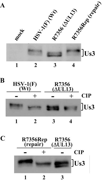
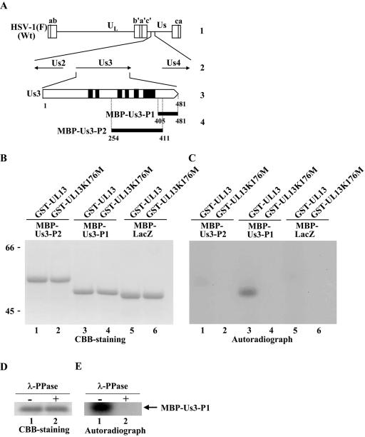
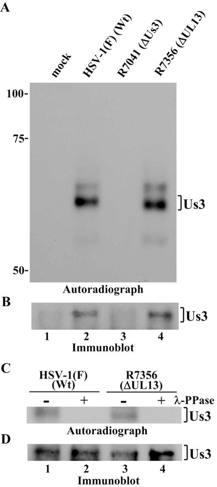
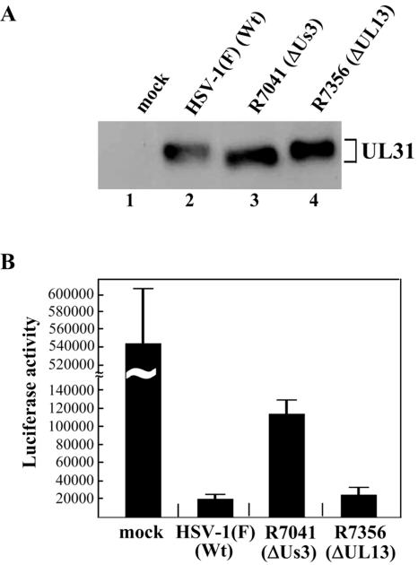
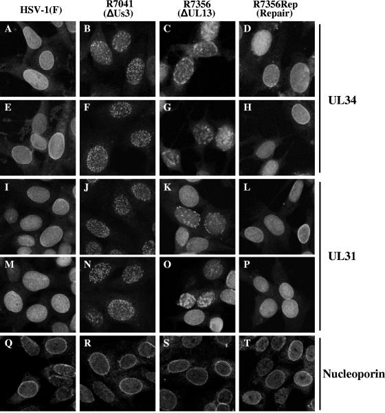
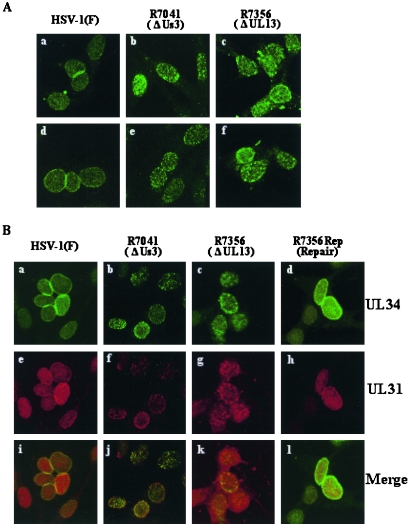
UL13 and Us3 are protein kinases encoded by herpes simplex virus 1. We report here that Us3 is a physiological substrate for UL13 in infected cells, based on the following observations. (i) The electrophoretic mobility, in denaturing gels, of Us3 isoforms from Vero cells infected with wild-type virus was slower than that of isoforms from cells infected with a UL13 deletion mutant virus (DeltaUL13). After treatment with phosphatase, the electrophoretic mobility of the Us3 isoforms from cells infected with wild-type virus changed, with one isoform migrating as fast as one of the Us3 isoforms from DeltaUL13-infected cells. (ii) A recombinant protein containing a domain of Us3 was phosphorylated by UL13 in vitro. (iii) The phenotype of DeltaUL13 resembles that of a recombinant virus lacking the Us3 gene (DeltaUs3) with respect to localization of the viral envelopment factors UL34 and UL31, whose localization has been shown to be regulated by Us3. UL34 and UL31 are localized in a smooth pattern throughout the nuclei of cells infected with wild-type virus, whereas their localization in DeltaUL13- and DeltaUs3-infected cells appeared as nuclear punctate patterns. These results indicate that UL13 phosphorylates Us3 in infected cells and regulates UL34 and UL31 localization, either by phosphorylating Us3 or by a Us3-independent mechanism.
Figures
Similar articles
-
UL13 protein kinase of herpes simplex virus 1 complexes with glycoprotein E and mediates the phosphorylation of the viral Fc receptor: glycoproteins E and I.Virology. 1998 Feb 1;241(1):37-48. doi: 10.1006/viro.1997.8963. Virology. 1998. PMID: 9454715
-
The UL13 and US3 Protein Kinases of Herpes Simplex Virus 1 Cooperate to Promote the Assembly and Release of Mature, Infectious Virions.PLoS One. 2015 Jun 26;10(6):e0131420. doi: 10.1371/journal.pone.0131420. eCollection 2015. PLoS One. 2015. PMID: 26115119 Free PMC article.
-
Herpes simplex virus protein kinases US3 and UL13 modulate VP11/12 phosphorylation, virion packaging, and phosphatidylinositol 3-kinase/Akt signaling activity.J Virol. 2014 Jul;88(13):7379-88. doi: 10.1128/JVI.00712-14. Epub 2014 Apr 16. J Virol. 2014. PMID: 24741093 Free PMC article.
-
Us3 Protein Kinase Encoded by HSV: The Precise Function and Mechanism on Viral Life Cycle.Adv Exp Med Biol. 2018;1045:45-62. doi: 10.1007/978-981-10-7230-7_3. Adv Exp Med Biol. 2018. PMID: 29896662 Review.
-
[Molecular mechanism by which Us3 protein kinase regulates the pathogenicity of herpes simplex virus type-1].Uirusu. 2016;66(1):83-90. doi: 10.2222/jsv.66.83. Uirusu. 2016. PMID: 28484184 Review. Japanese.
Cited by
-
Characterization of the subcellular localization of herpes simplex virus type 1 proteins in living cells.Med Microbiol Immunol. 2011 Feb;200(1):61-8. doi: 10.1007/s00430-010-0175-9. Epub 2010 Oct 15. Med Microbiol Immunol. 2011. PMID: 20949280
-
Conserved Herpesvirus Protein Kinases Target SAMHD1 to Facilitate Virus Replication.Cell Rep. 2019 Jul 9;28(2):449-459.e5. doi: 10.1016/j.celrep.2019.04.020. Cell Rep. 2019. PMID: 31291580 Free PMC article.
-
VHS, US3 and UL13 viral tegument proteins are required for Herpes Simplex Virus-Induced modification of protein kinase R.Sci Rep. 2020 Mar 27;10(1):5580. doi: 10.1038/s41598-020-62619-2. Sci Rep. 2020. PMID: 32221365 Free PMC article.
-
Characterization of a Herpes Simplex Virus 1 (HSV-1) Chimera in Which the Us3 Protein Kinase Gene Is Replaced with the HSV-2 Us3 Gene.J Virol. 2015 Oct 21;90(1):457-73. doi: 10.1128/JVI.02376-15. Print 2016 Jan 1. J Virol. 2015. PMID: 26491159 Free PMC article.
-
Reconstitution of herpes simplex virus type 1 nuclear capsid egress in vitro.J Virol. 2006 Oct;80(19):9741-53. doi: 10.1128/JVI.00061-06. J Virol. 2006. PMID: 16973578 Free PMC article.
References
-
- Advani, S. J., R. Brandimarti, R. R. Weichselbaum, and B. Roizman. 2000. The disappearance of cyclins A and B and the increase in activity of the G2/M-phase cellular kinase cdc2 in herpes simplex virus 1-infected cells require expression of the alpha22/U(S)1.5 and U(L)13 viral genes. J. Virol. 74:8-15. - PMC - PubMed
-
- Asano, S., T. Honda, F. Goshima, D. Watanabe, Y. Miyake, Y. Sugiura, and Y. Nishiyama. 1999. US3 protein kinase of herpes simplex virus type 2 plays a role in protecting corneal epithelial cells from apoptosis in infected mice. J. Gen. Virol. 80:51-56. - PubMed
Publication types
MeSH terms
Substances
LinkOut - more resources
Full Text Sources
Research Materials
