. 2006 Feb;80(3):1497-512.
doi: 10.1128/JVI.80.3.1497-1512.2006.

The cellular localization pattern of Varicella-Zoster virus ORF29p is influenced by proteasome-mediated degradation

Affiliations

The cellular localization pattern of Varicella-Zoster virus ORF29p is influenced by proteasome-mediated degradation

Christina L Stallings et al. J Virol. 2006 Feb.

Abstract

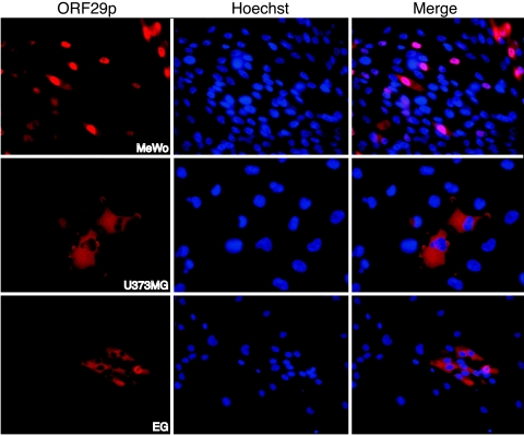
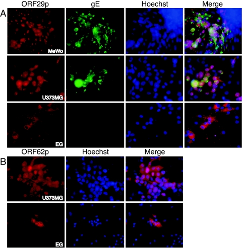
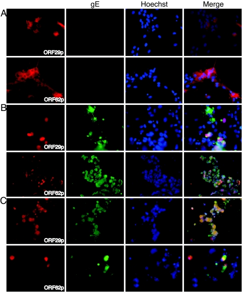
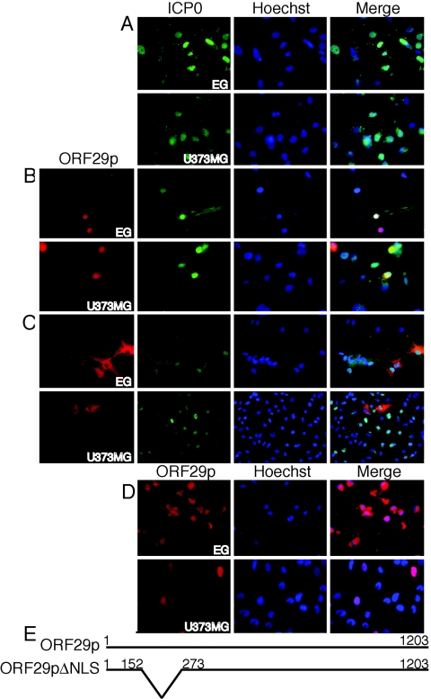
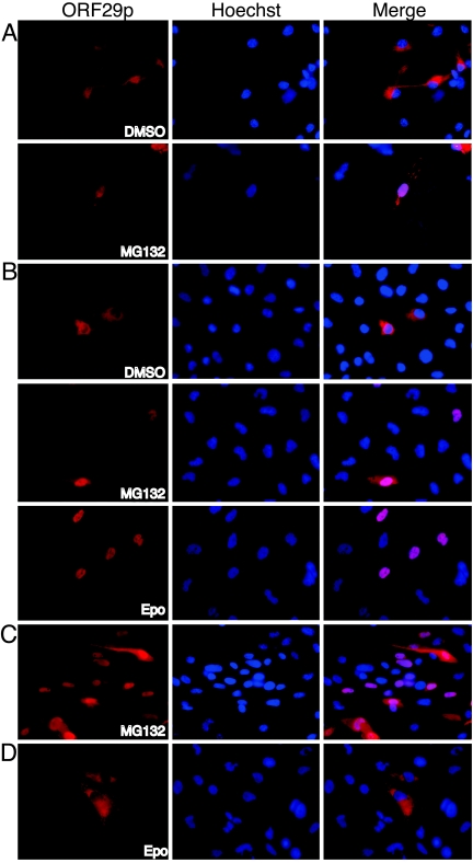
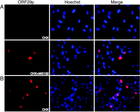
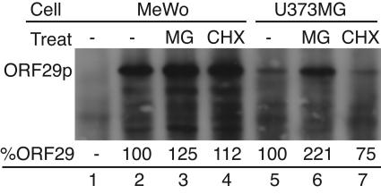
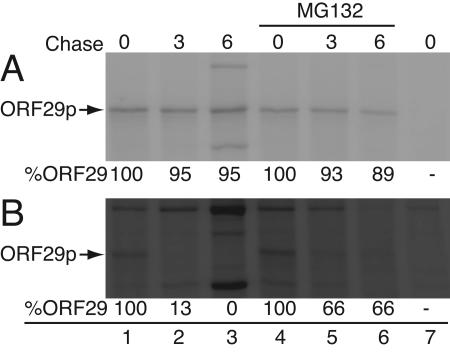
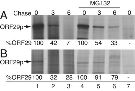
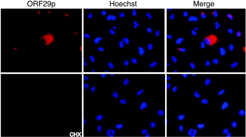
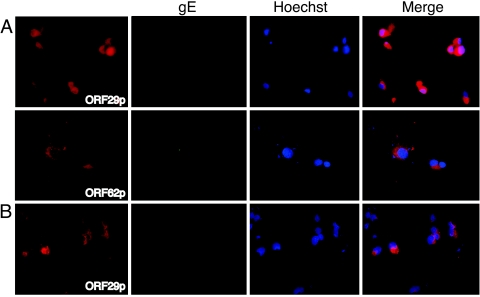
Varicella-zoster virus (VZV) open reading frame 29 (ORF29) encodes a single-stranded DNA binding protein. During lytic infection, ORF29p is localized primarily to infected-cell nuclei, whereas during latency it appears in the cytoplasm of infected neurons. Following reactivation, ORF29p accumulates in the nucleus. In this report, we analyze the cellular localization patterns of ORF29p during VZV infection and during autonomous expression. Our results demonstrate that ORF29p is excluded from the nucleus in a cell-type-specific manner and that its cellular localization pattern may be altered by subsequent expression of VZV ORF61p or herpes simplex virus type 1 ICP0. In these cases, ORF61p and ICP0 induce nuclear accumulation of ORF29p in cell lines where it normally remains cytoplasmic. One cellular system utilized by ICP0 to influence protein abundance is the proteasome degradation pathway. Inhibition of the 26S proteasome, but not heat shock treatment, resulted in accumulation of ORF29p in the nucleus, similar to the effect of ICP0 expression. Immunofluorescence microscopy and pulse-chase experiments reveal that stabilization of ORF29p correlates with its nuclear accumulation and is dependent on a functional nuclear localization signal. ORF29p nuclear translocation in cultured enteric neurons and cells derived from an astrocytoma is reversible, as the protein's distribution and stability revert to the previous states when the proteasomal activity is restored. Thus, stabilization of ORF29p leads to its nuclear accumulation. Although proteasome inhibition induces ORF29p nuclear accumulation, this is not sufficient to reactivate latent VZV or target the immediate-early protein ORF62p to the nucleus in cultured guinea pig enteric neurons.


Figures

FIG. 1.
FIG. 1.
Cellular localization of ORF29p. Cultured guinea pig EG, MeWo, and U373MG cells were infected with AdORF29 at an MOI of 50. The localization of ORF29p was examined at 5 dpi by indirect immunofluorescence microscopy. Nuclei were counterstained with Hoechst 33258.
FIG. 2.
FIG. 2.
Localization of virus-specified proteins in cells infected with cell-free VZV. Cultured EG, MeWo, and U373MG cells were infected with cell-free VZV at an MOI of 1. The expression and distribution of ORF29p and gE (A) and ORF62p (B) were examined at 3 dpi by indirect immunofluorescence microscopy.
FIG. 3.
FIG. 3.
Localization of viral proteins in VZV-infected guinea pig EG. Cultured EG were infected with cell-free VZV at an MOI of 1. At 3 dpi, cells were superinfected with either Ad.MmCMV (A), MLP-0 (B), or AdORF61 (C) at an MOI of 50 for 48 h prior to analysis by indirect immunofluorescence. ORF29p, ORF62p, and gE were detected after incubation with specific antisera.
FIG. 4.
FIG. 4.
Localization of ORF29p in cells expressing ORF29p and HSV-1 ICP0 or VZV ORF61p. (A) Cultured EG and U373MG cells were infected with MLP-0 at an MOI of 50. ICP0 was detected at 5 dpi by indirect immunofluorescence microscopy after incubation with specific antiserum. (B to D) EG and U373MG cells were also infected with either an adenovirus expressing the wild-type ORF29p (AdORF29) (B and D) or an adenovirus expressing a mutant ORF29p with a deletion in the NLS (AdORF29ΔNLS) (C) at an MOI of 50. After 72 h, the EG and U373MG cells were superinfected with MLP-0 (B and C) or AdORF61 (D) at an MOI of 50. The cultures were incubated for another 48 h before the distributions of ORF29p, ORF29pΔNLS, and ICP0 were examined by indirect immunofluorescence microscopy. (E) Schematic of the amino acids deleted from the ORF29pΔNLS mutant.
FIG. 5.
FIG. 5.
Localization of ORF29p in cells after sequential expression of HSV-1 ICP0 insertion mutants. (A) U373MG cells were infected with AdORF29 at an MOI of 50. After 72 h, the cells were superinfected with either Ad-0/125 or Ad-0/88 at an MOI of 50. The cultures were incubated for another 48 h before the distributions of ORF29p and ICP0 variants were examined by indirect immunofluorescence microscopy. (B) Clustal X alignment of HSV-1 ICP0 (accession no. P08393) and VZV ORF61p (accession no. P09309) amino acid sequences. The underlines mark the functional domains as characterized in ICP0. The red underline denotes the RING finger motif encoded by exon 2 that possesses E3 ubiquitin ligase activity as well as binds the cdc34 E2 ubiquitin-conjugating enzyme; this domain is well conserved between the two proteins. The black underline identifies the domain that confers E3 ubiquitin ligase activity on HSV-1 ICP0; note that this domain is not well conserved between these proteins. The asterisks identify positions that have a single, fully conserved residue, the colons identify highly conserved residues, and the periods identify weakly conserved residues at the indicated positions.
FIG. 6.
FIG. 6.
Localization of ORF29p in cells treated with proteasome inhibitors. (A to C) Cultured EG (A), U373MG (B), and MeWo (C) cells grown on glass coverslips were infected with AdORF29 at an MOI of 50. (D) U373MG cells were infected with AdORF29ΔNLS at an MOI of 50. At 3 dpi, infected cultures were treated with either DMSO, 20 μM MG132, or 20 μM epoxomicin (Epo) for 6 h prior to analysis. ORF29p and ORF29pΔNLS were detected by indirect immunofluorescence microscopy after incubation with antiserum specific for ORF29p.
FIG. 7.
FIG. 7.
Localization of ORF29p in cells treated with cycloheximide. U373MG (A) and MeWo (B) cells grown on glass coverslips were infected with AdORF29 at an MOI of 50. At 3 dpi, infected cultures were treated with 50 μg/ml of cycloheximide (CHX) or with 50 μg/ml cycloheximide and 20 μM MG132 for 6 h and fixed. ORF29p was detected by indirect immunofluorescence microscopy after reaction with specific antisera.
FIG. 8.
FIG. 8.
Western blot analysis of ORF29p after treatment with MG132 or cycloheximide. MeWo and U373MG cells were infected with AdORF29 at an MOI of 50 (lanes 2 to 7) or mock infected (lane 1). At 3 dpi, cells were treated with either 20 μM MG132 (MG) (lanes 3 and 6), 50 μg/ml cycloheximide (CHX) (lanes 4 and 7), or DMSO (lanes 1, 2, and 5) for 6 h. Cells were harvested, lysed in RIPA buffer, and subjected to SDS-PAGE on an 8% gel. Proteins were transferred to a nitrocellulose membrane, reacted with specific antisera, and visualized using an horseradish peroxidase reporter substrate. Band intensity (percent ORF29) was quantified relative to ORF29p levels in the DMSO-treated sample by using the ImageJ program, and total protein levels per well were normalized against a band that represents a cross-reacting protein that was detected in all lanes.
FIG. 9.
FIG. 9.
Autoradiograph of metabolically labeled ORF29p. MeWo (A) and U373MG (B) cells were infected with AdORF29 at an MOI of 50 (lanes 1 to 6) or mock infected (lane 7). At 3 dpi, cells were labeled with 500 μCi/ml Tran35S-label for 1 h in the presence of either 20 μM MG132 (lanes 4 to 6) or DMSO (lanes 1 to 3 and 7). The labeling medium was replaced with DMEM supplemented with 10% fetal bovine serum and either 20 μM MG132 (lanes 4 to 6) or DMSO (lanes 1 to 3 and 7) for the indicated chase times. Cells were harvested and lysed in RIPA buffer, and ORF29p was immunoprecipitated and subjected to analysis by SDS-PAGE on an 8% gel. Proteins were visualized by autoradiography. Band intensity was quantified using the ImageJ program, and total protein levels per well were normalized as described in the legend to Fig. 8. Percent ORF29 was calculated relative to the amount present at the 0-h chase time point.
FIG. 10.
FIG. 10.
Autoradiograph of metabolically labeled ORF29pΔNLS. MeWo (A) and U373MG (B) cells were infected with AdORF29ΔNLS at an MOI of 50 (lanes 1 to 6) or mock infected (lane 7). At 3 dpi, cells were labeled with 500 μCi/ml Tran35S-label for 1 h in the presence of either 20 μM MG132 (lanes 4 to 6) or DMSO (lanes 1 to 3 and 7). The labeling medium was replaced with DMEM supplemented with 10% fetal bovine serum and either 20 μM MG132 (lanes 4 to 6) or DMSO (lanes 1 to 3 and 7) for the indicated chase time. Cells were harvested, lysed in RIPA buffer, immunoprecipitated with ORF29p-specific antiserum, and subjected to SDS-PAGE analysis on an 8% gel. Proteins were visualized by autoradiography. Band intensity was quantified using the ImageJ program, and total protein levels per well were normalized against a nonspecific band detected in all lanes. Percent ORF29pΔNLS (%ORF29) was calculated relative to the amount present at the 0-h chase time point.
FIG. 11.
FIG. 11.
Localization of ORF29p in U373MG cells after MG132 reversal. U373MG cells grown on coverslips were infected with AdORF29 at an MOI of 50. At 3 dpi, infected cultures were treated with 20 μM MG132. After 6 h, the cells were washed three times in PBS and the medium was replaced with either DMEM supplemented with 10% fetal bovine serum or DMEM supplemented with 10% fetal bovine serum and 50 μg/ml of cycloheximide (CHX) for 6 h and then fixed in situ. ORF29p was detected by indirect immunofluorescence microscopy after incubation with specific antisera.
FIG. 12.
FIG. 12.
Localization of viral proteins in VZV-infected guinea pig EG treated with proteasome inhibitors. Cultured EG were infected with cell-free VZV at an MOI of 1. At 3 dpi, cells were treated with 20 μM MG132 for 24 h prior to fixing onto glass slides (A) or for 6 h before washing three times in PBS (B) and incubating in normal maintenance medium for an additional 18 h before processing. ORF29p, ORF62p, and gE were detected by indirect immunofluorescence microscopy after incubation with specific antisera.

Similar articles

Cited by

References

    1. Arvin, A. M. 1996. Varicella-zoster virus. Clin. Microbiol. Rev. 9:361-381. - PMC - PubMed
    1. Assouline, J. G., M. J. Levin, E. O. Major, B. Forghani, S. E. Straus, and J. M. Ostrove. 1990. Varicella-zoster virus infection of human astrocytes, Schwann cells, and neurons. Virology 179:834-844. - PubMed
    1. Boucaud, D., H. Yoshitake, J. Hay, and W. Ruyechan. 1998. The varicella-zoster virus (VZV) open-reading frame 29 protein acts as a modulator of a late VZV gene promoter. J Infect. Dis. 178(Suppl. 1):S34-S38. - PubMed
    1. Boutell, C., A. Orr, and R. D. Everett. 2003. PML residue lysine 160 is required for the degradation of PML induced by herpes simplex virus type 1 regulatory protein ICP0. J. Virol. 77:8686-8694. - PMC - PubMed
    1. Boutell, C., S. Sadis, and R. D. Everett. 2002. Herpes simplex virus type 1 immediate-early protein ICP0 and its isolated RING finger domain act as ubiquitin E3 ligases in vitro. J. Virol. 76:841-850. - PMC - PubMed

Publication types

MeSH terms

LinkOut - more resources